

Neutralizing Antibodies Against Allosteric Proteins: Insights From a Bacterial Adhesin
- PMID: 35798162
- PMCID: PMC9398984
- DOI: 10.1016/j.jmb.2022.167717
Neutralizing Antibodies Against Allosteric Proteins: Insights From a Bacterial Adhesin
Abstract
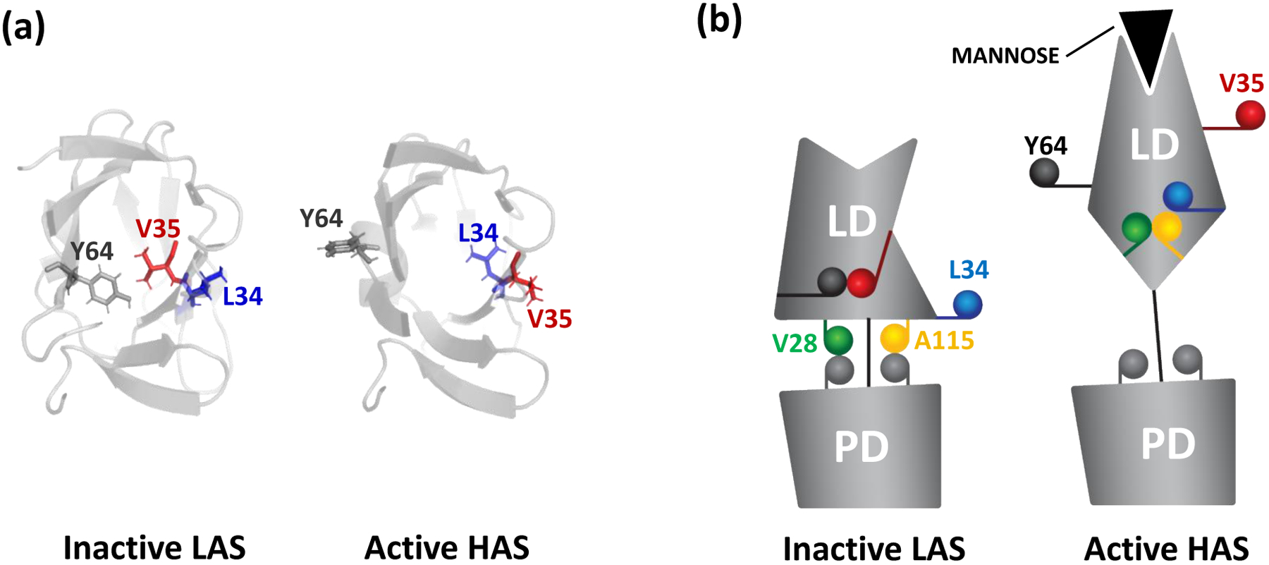
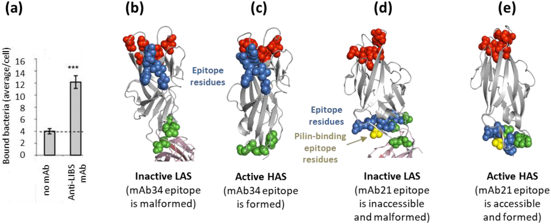
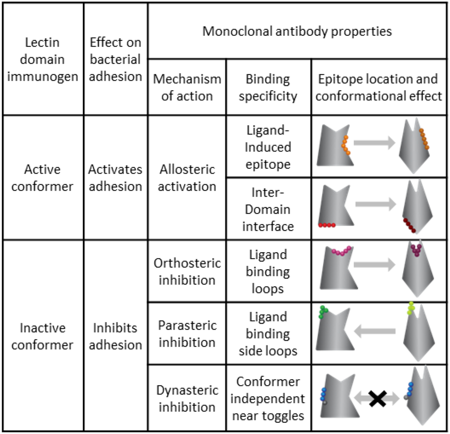
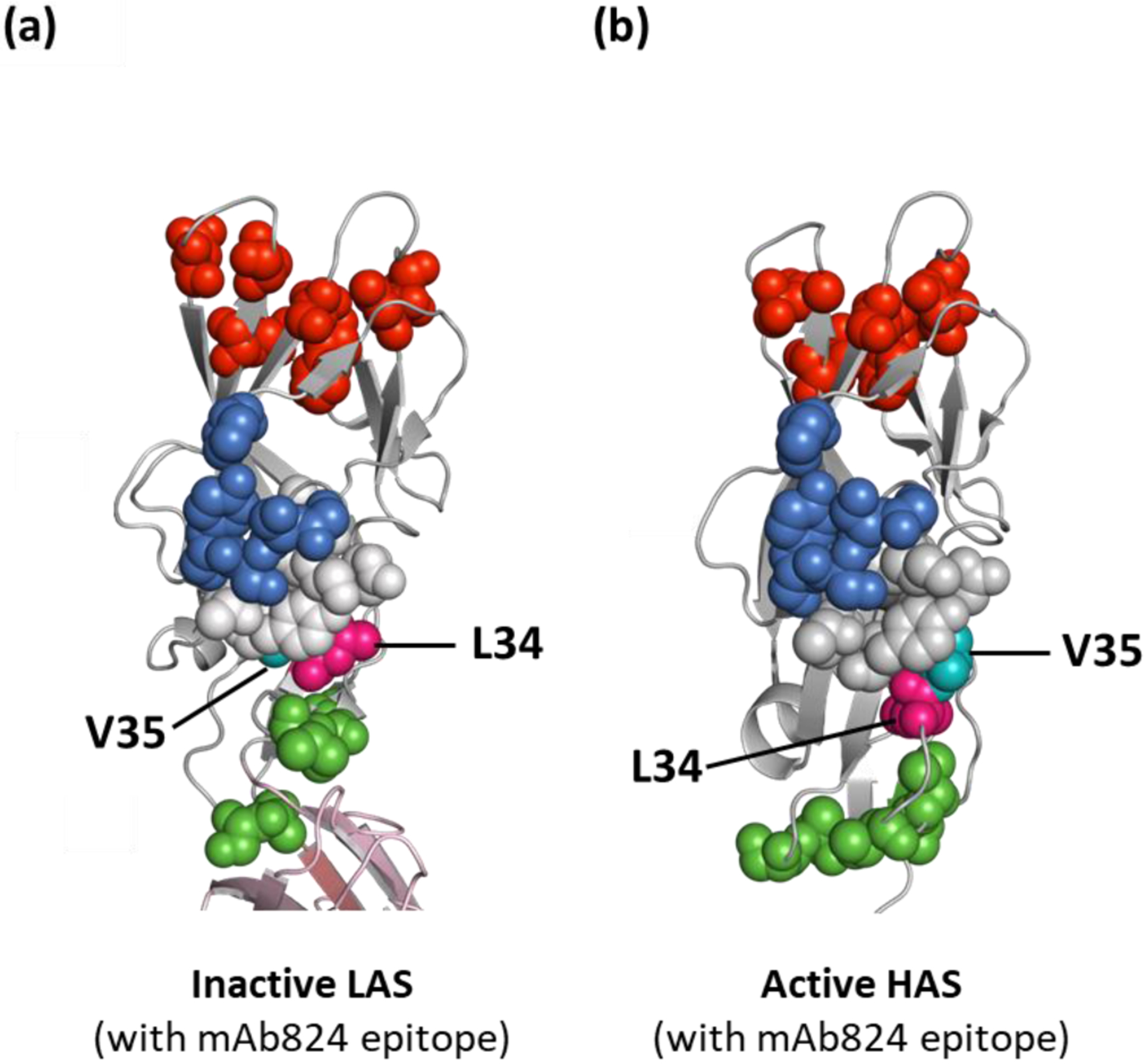
Allosteric proteins transition between 'inactive' and 'active' states. In general, such proteins assume distinct conformational states at the level of secondary, tertiary and/or quaternary structure. Different conformers of an allosteric protein can be antigenically dissimilar and induce antibodies with a highly distinctive specificities and neutralizing functional effects. Here we summarize studies on various functional types of monoclonal antibodies obtained against different allosteric conformers of the mannose-specific bacterial adhesin FimH - the most common cell attachment protein of Escherichia coli and other enterobacterial pathogens. Included are types of antibodies that activate the FimH function via interaction with ligand-induced binding sites or by wedging between domains as well as antibodies that inhibit FimH through orthosteric, parasteric, or novel dynasteric mechanisms. Understanding the molecular mechanism of antibody action against allosteric proteins provides insights on how to design antibodies with a desired functional effect, including those with neutralizing activity against bacterial and viral cell attachment proteins.
Keywords: FimH; adhesin; attachment; conformation; inhibition.
Copyright © 2022 Elsevier Ltd. All rights reserved.
Conflict of interest statement
Declaration of Competing Interest The authors declare that they have no known competing financial interests or personal relationships that could have appeared to influence the work reported in this paper.
Figures


Similar articles
-
Allosteric coupling in the bacterial adhesive protein FimH.J Biol Chem. 2013 Aug 16;288(33):24128-39. doi: 10.1074/jbc.M113.461376. Epub 2013 Jul 2. J Biol Chem. 2013. PMID: 23821547 Free PMC article.
-
Recombinant FimH Adhesin Demonstrates How the Allosteric Catch Bond Mechanism Can Support Fast and Strong Bacterial Attachment in the Absence of Shear.J Mol Biol. 2022 Sep 15;434(17):167681. doi: 10.1016/j.jmb.2022.167681. Epub 2022 Jun 11. J Mol Biol. 2022. PMID: 35697293 Free PMC article.
-
Type 1 fimbrial adhesin FimH elicits an immune response that enhances cell adhesion of Escherichia coli.Infect Immun. 2011 Oct;79(10):3895-904. doi: 10.1128/IAI.05169-11. Epub 2011 Jul 18. Infect Immun. 2011. PMID: 21768279 Free PMC article.
-
Molecular structure of adhesin domains in Escherichia coli fimbriae.Int J Med Microbiol. 2005 Oct;295(6-7):479-86. doi: 10.1016/j.ijmm.2005.06.010. Int J Med Microbiol. 2005. PMID: 16238022 Review.
-
Targeting Dynamical Binding Processes in the Design of Non-Antibiotic Anti-Adhesives by Molecular Simulation-The Example of FimH.Molecules. 2018 Jul 5;23(7):1641. doi: 10.3390/molecules23071641. Molecules. 2018. PMID: 29976867 Free PMC article. Review.
Cited by
-
Antibodies disrupt bacterial adhesion by ligand mimicry and allosteric interference.bioRxiv [Preprint]. 2024 Dec 14:2024.12.06.627246. doi: 10.1101/2024.12.06.627246. bioRxiv. 2024. PMID: 39713463 Free PMC article. Preprint.
-
Recent advances in surface plasmon resonance for the detection of ovarian cancer biomarkers: a thorough review.Mikrochim Acta. 2024 Oct 9;191(11):659. doi: 10.1007/s00604-024-06740-3. Mikrochim Acta. 2024. PMID: 39382786 Review.
-
Beyond Antibiotics: What the Future Holds.Antibiotics (Basel). 2024 Sep 25;13(10):919. doi: 10.3390/antibiotics13100919. Antibiotics (Basel). 2024. PMID: 39452186 Free PMC article. Review.
-
Effect of Gravity on Bacterial Adhesion to Heterogeneous Surfaces.Pathogens. 2023 Jul 15;12(7):941. doi: 10.3390/pathogens12070941. Pathogens. 2023. PMID: 37513788 Free PMC article.
-
Structure-based design of an immunogenic, conformationally stabilized FimH antigen for a urinary tract infection vaccine.PLoS Pathog. 2025 Feb 19;21(2):e1012325. doi: 10.1371/journal.ppat.1012325. eCollection 2025 Feb. PLoS Pathog. 2025. PMID: 39970181 Free PMC article.
References
-
- Monod J, Chance and Necessity: An Essay on the Natural Philosophy of Modern Biology, Vintage Books, 1971.
-
- Monod J, Wyman J, Changeux JP, On the Nature of Allosteric Transitions: a Plausible Model, J Mol Biol. 12 (1965) 88–118. http://www.ncbi.nlm.nih.gov/entrez/query.fcgi?cmd=Retrieve&db=PubMed&dop.... - PubMed
Publication types
MeSH terms
Substances
Grants and funding
LinkOut - more resources
Full Text Sources
