. 2022 Sep:205:105373.
doi: 10.1016/j.antiviral.2022.105373. Epub 2022 Jul 4.

Hydroxypropyl-beta-cyclodextrin (HP-BCD) inhibits SARS-CoV-2 replication and virus-induced inflammatory cytokines

Affiliations

Hydroxypropyl-beta-cyclodextrin (HP-BCD) inhibits SARS-CoV-2 replication and virus-induced inflammatory cytokines

Bruno Braz Bezerra et al. Antiviral Res. 2022 Sep.

Abstract

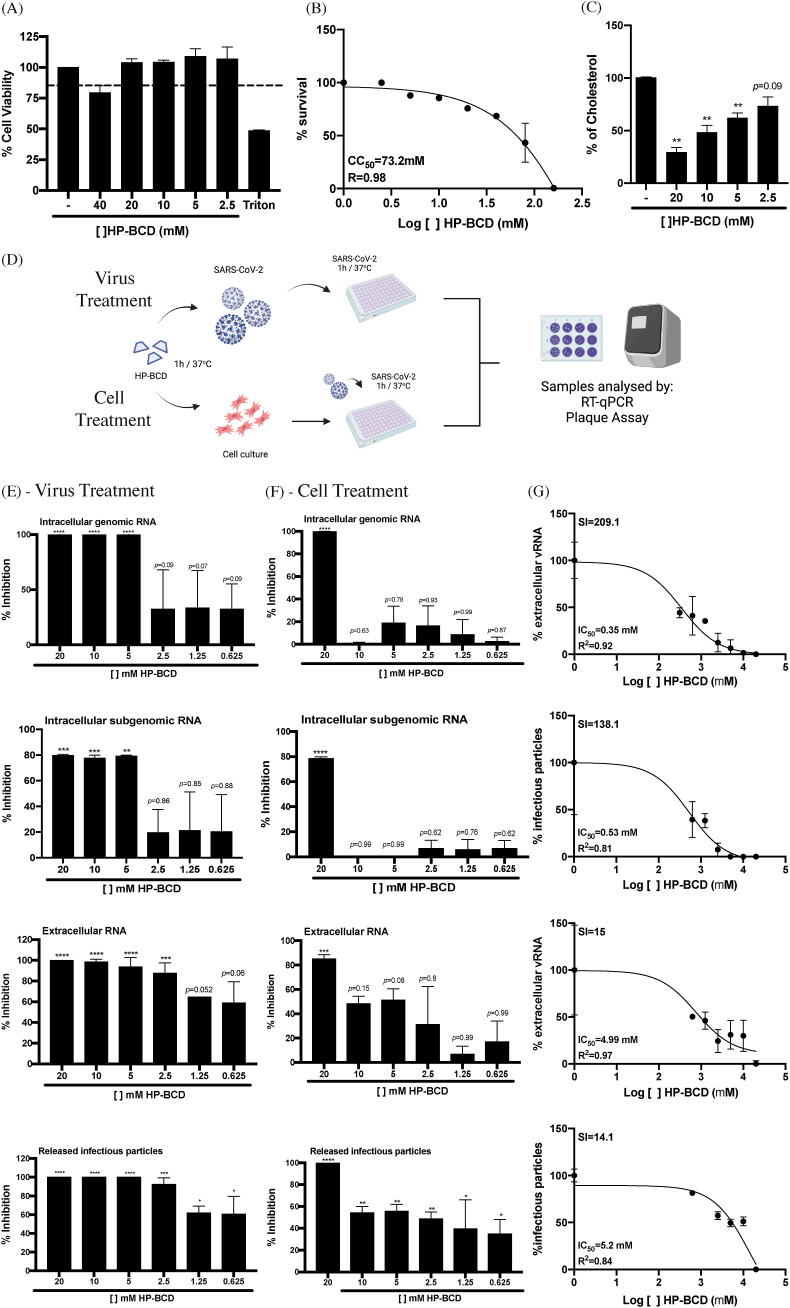
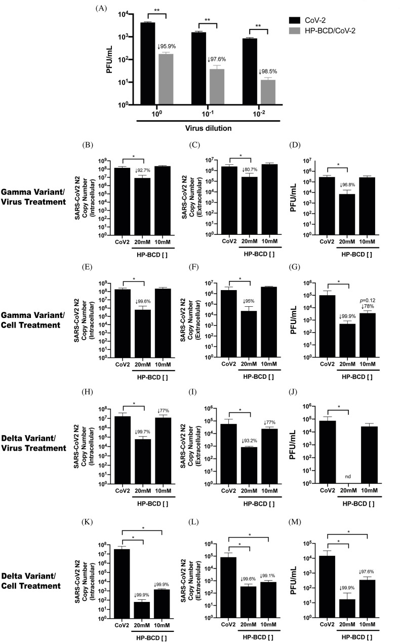
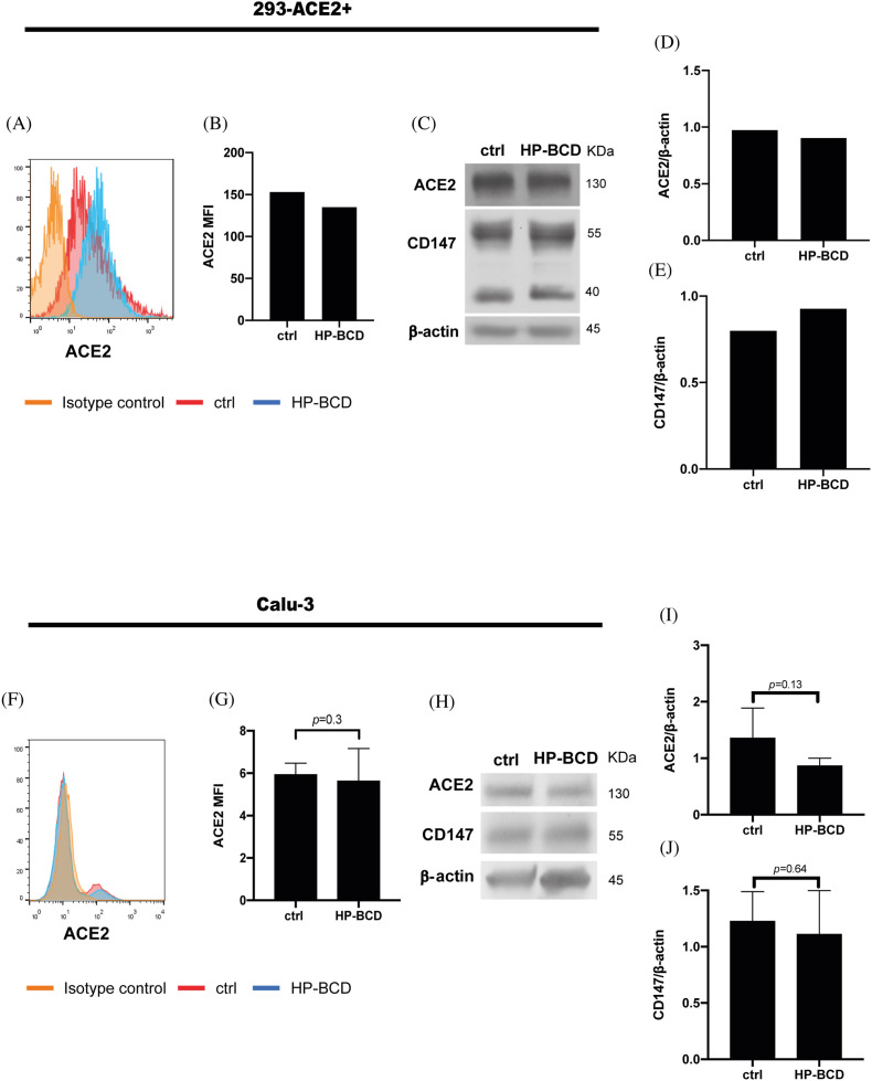
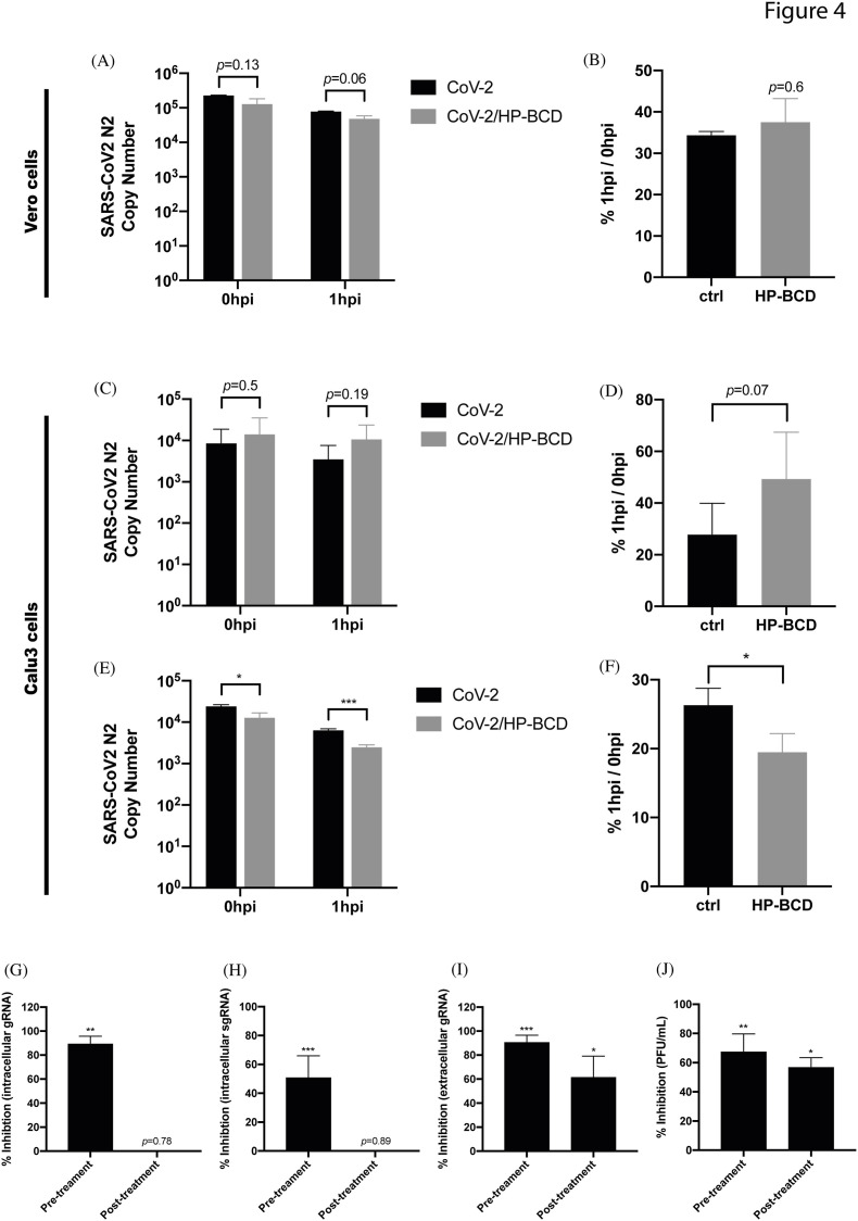
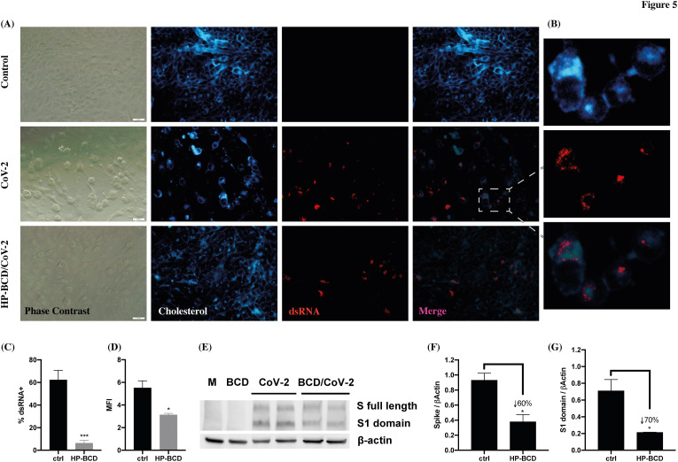
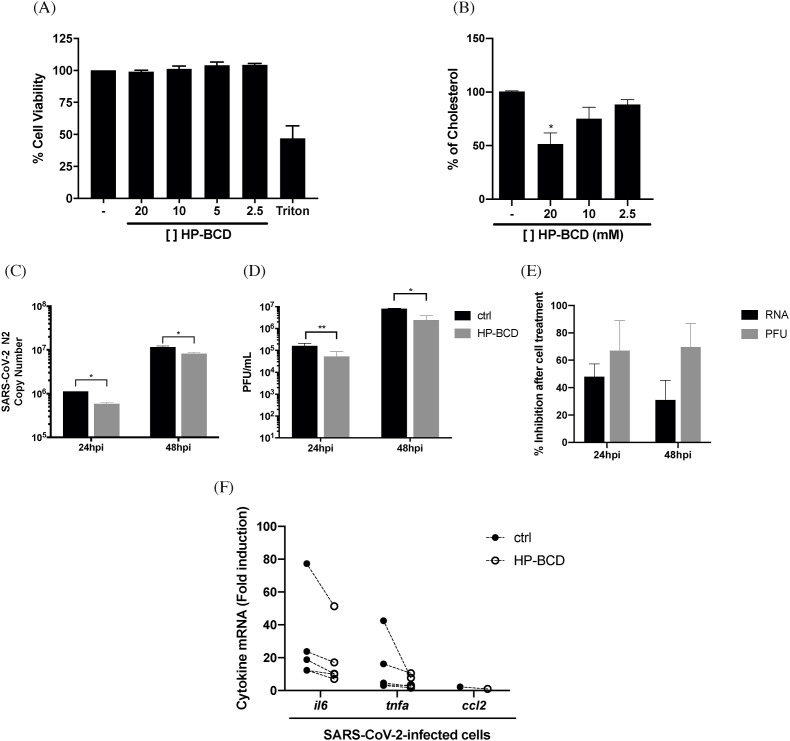
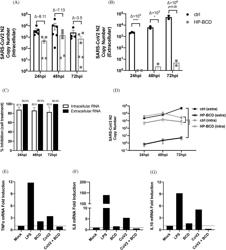
COVID-19 is marked by extensive damage to the respiratory system, often accompanied by systemic manifestations, due to both viral cytopathic effects and hyperinflammatory syndrome. Therefore, the development of new therapeutic strategies or drug repurposing aiming to control virus replication and inflammation are required to mitigate the impact of the disease. Hydroxypropyl-beta-cyclodextrin (HP-BCD) is a cholesterol-sequestering agent with antiviral activity that has been demonstrated against enveloped viruses in in vitro and in vivo experimental models. We also demonstrated that HP-BCD has an immunomodulatory effect, inhibiting the production of selected proinflammatory cytokines induced by microbial products. Importantly, this drug has been used in humans for decades as an excipient in drug delivery systems and as a therapeutic agent in the treatment of Niemann pick C disease. The safety profile for this compound is well established. Here, we investigated whether HP-BCD would affect SARS-CoV-2 replication and virus-induced inflammatory response, using established cell lines and primary human cells. Treating virus or cells with HP-BCD significantly inhibited SARS-CoV-2 replication with a high selective index. A broad activity against distinct SARS-CoV-2 variants was evidenced by a remarkable reduction in the release of infectious particles. The drug did not alter ACE2 surface expression, but affected cholesterol accumulation into intracellular replication complexes, lowering virus RNA and protein levels, and reducing virus-induced cytopathic effects. Virus replication was also impaired by HP-BCD in Calu-3 pulmonary cell line and human primary monocytes, in which not only the virus, but also the production of proinflammatory cytokines were significantly inhibited. Given the pathophysiology of COVID-19 disease, these data indicate that the use HP-BCD, which inhibits both SARS-CoV2 replication and production of proinflammatory cytokines, as a potential COVID-19 therapeutic warrants further investigation.

Keywords: Beta-cyclodextrin; COVID-19; Cholesterol; Inflammation; SARS-COV-2; Virus replication.


Conflict of interest statement

The authors declare that they have no known competing financial interests or personal relationships that could have appeared to influence the work reported in this paper.

Figures

Fig. 1
Fig. 1
HP-BCD inhibits SARS-CoV-2 replication in Vero cells. A-C) Vero cells were treated with the indicated concentrations of HP-BCD for 1h, the cells were washed and then cultured in complete culture medium. A-B) After 72 h, cell viability was measured using cell-titer blue kit (A); the data was analyzed and the CC50 was calculated using GraphPad Prisma software (B). C) Total cholesterol concentration was measured by amplex red reagent after 1 h treatment. D) Schematic representation of the following assays. Top (Virus treatment): SARS-CoV-2 stock samples (A2 variant) were treated or not with HP-BCD for 1 h, and then inoculated in Vero cells at a MOI of 0.1. After 1 h of virus adsorption, the cells were washed and cultured in HP-BCD-free culture medium for additional 72 h. Bottom (Cell treatment): Vero cells were treated with HP-BCD for 1 h, the cells were washed, and cultured in HP-BCD-free culture medium; then, virus was inoculated at a MOI of 0.1, as described. This figure was created with BioRender.com. E, F) SARS-CoV-2 stock samples or Vero cells were treated with HP-BCD and the infection proceeded, as indicated in (D). After 72 hpi, the cell lysates and culture supernatants were harvested. The concentration of intracellular genomic and subgenomic virus RNA, and released genomic RNA were measured by RT-qPCR; titration of released infectious virus particles was performed by plaque assay. The bars indicate the percentage inhibition of RNA copy numbers or PFU/ml, in relation to untreated virus or cells, obtained from five independent experiments. Statistical analyses were performed by one-way anova, followed by Dunnet's multiple comparison tests; * represents p < 0.05; **p < 0.01; ***p < 0.001; ****p < 0.0001. G, H) The released virus RNA and infectious particles data obtained after virus treatment (G) or cell treatment (H) were analyzed, and the IC50 was calculated using GraphPad Prisma software. The selectivity indexes (SI) were calculated using the CC50 data obtained in (B).
Fig. 2
Fig. 2
HP-BCD presents a broad antiviral activity against different SARS-CoV-2 variants of concern. A) SARS-COV-2 stock samples were incubated or not with HP-BCD for 1 h, serially diluted and inoculated onto Vero E6 cells for virus titration. After 48 h, virus titers were calculated and indicated as PFU/ml. Statistical analysis of two independent experiments were performed using t-test; ** represents p < 0.01, and insert numbers indicate the percentage inhibition in relation to non treated viruses. B-M) Vero E6 cells were infected with SARS-CoV-2 sequenced and characterized as Gamma (BG) or Delta (HM) variants, at a MOI of 0.1. Cell-free virus or host cell treatments were performed as described in Fig. 1. After 72 h, cell lysates and culture supernatants were harvested. The concentration of intracellular (B, E, H, K) and released virus RNA (N2 copy numbers) (C, F, I, L) were measured by RT-qPCR; titration of released infectious virus particles was performed by plaque assay and represented as PFU/ml (D, G, J, M). The bars indicate the average and SD of two independent experiments, in triplicate. Statistical analyses were performed by one-way anova, followed by Dunnet's multiple comparison; * represents p < 0.05, and insert numbers indicate the percentage inhibition in relation to infected non treated cells; nd-not detected.
Fig. 3
Fig. 3
HP-BCD does not affect the expression of SARS-CoV-2 receptors. ACE-2-expressing HEK-293 cells or Calu-3 cells were treated with 20 mM HP-BCD for 1 h. The expression of ACE2 in intact nonpermeabilized cells was analyzed by flow cytometry, whereas total expression of ACE2 and CD147 were evaluated in the cell lysates by western blotting. Representative histograms of ACE2 staining and flow cytometry analysis are depicted in (A) and (F); and the MFI average obtained from independent experiments represented in (B) and (G). For the western blotting analysis, the membranes were stained with anti-ACE2, anti-CD147, and anti-β-actin, as a loading control. Representative blots are demonstrated in (C) and (H); the expression level of ACE-2 (D, I) and CD147 (E, J) were estimated using ImageJ software and normalized according to β-actin expression. Three experiments were run with Calu-3 cells and statistical analyses were performed by paired t-test.
Fig. 4
Fig. 4
SARS-CoV-2 incubation, but not cell treatment, with HP-BCD reduces SARS-CoV-2 adsorption and entry. A–B) Vero cells were treated with 20 mM HP-BCD for 1h, washed, and cultured in HP-BCD-free culture medium. SARS-CoV-2 (A2 strain) was inoculated at an MOI of 0.1 for 1 h/4 °C for virus adsorption and harvested (0 hpi) or further incubated for 1 h/37 °C for virus entry (1 hpi). Virus RNA was measured by qRT-PCR (A) and the frequency of virus entry in relation to virus adsorption (%1 hpi/0 hpi) was calculated (B). C-D) Calu-3 cells were treated with HP-BCD, infected with SARS-CoV2, and virus adsorption and entry were analyzed as in (AB). E-F) SARS-CoV-2 stock samples were incubated with 20 mM HP-BCD and then inoculated onto Calu-3 cells cultures. Analysis of virus RNA copy numbers after virus adsorption (0 hpi) and virus entry (1 hpi) was measured as in (A, C) and the entrance ratio (1 hpi/0 hpi) was calculated as in (B, D). The bars indicate the average and SD from three independent experiments; statistical analyses were performed by unpaired t-test. G-J) Vero cells were treated or not with HP-BCD and infected with SARS-CoV-2 as previously described (pre-treatment). In some wells, the cells were incubated with SARS-CoV-2 for 1 h, for virus adsorption, washed, and maintained in culture for 1 h more. Then, the cells were treated with 20 mM HP-BCD (post treatment). After 48 hpi, the analysis of intracellular virus gRNA (G), sgRNA (H), and extracelular gRNA (I) were performed by RT-qPCR, and titration of released infectious particles (J) was performed by plaque assay. The bars indicate the percentage inhibition of RNA copy numbers or PFU/ml, in relation to untreated virus or cells, obtained from two independent experiments; statistical analyses were performed by one-way anova, followed by Dunnet's multiple comparison tests; * represents p < 0.05; **p < 0.01; ***p < 0.001.
Fig. 5
Fig. 5
HP-BCD treatment inhibits virus RNA and protein expression and reduces SARS-CoV-2-mediated CPE. Vero cells were treated with 20 mM HP-BCD for 1 h. The cells were washed, and mock-treated (control) or infected with SARS-CoV-2 (gamma isolate; MOI = 0.1) for 48 h. A-C) Expression and distribution of dsRNA and cholesterol were evaluated by staining the cells with J2 antibody and filipin, followed by fluorescence microscopy analysis. Representative images are demonstrated in (A; scale bar = 20 μm), with a zoom of infected cell depicted in (B); mean fluorescence intensity (MFI) and frequency of J2 positive staining (% dsRNA+) of 5 randomly selected fields, with a total of about 500 cells, were calculated using ImageJ software and are represented in (C) and (D), respectively. E-G) Calu-3 cells were lysed and the expression of Spike protein was analyzed by western blotting. A representative blot is demonstrated in (E) and the expression level of whole S (F) or S1 (G) was calculated based on β-actin loading control staining, using ImageJ software. Statistical analyses were performed by unpaired t-test; * represents p < 0.05; ***p < 0.001; insert numbers indicate the percentage inhibition in relation to untreated cells.
Fig. 6
Fig. 6
Treatment of Calu3 cells with HP-BCD inhibit SARS-CoV-2 replication and cytokine production. Calu-3 cells were treated with the indicated concentrations of HP-BCD for 1 h, the cells were washed and then cultured in HP-BCD-free culture medium. A) After 72 h, cell viability was measured using cell-titer blue kit. B) Total cholesterol concentration was measured after 1 h treatment by amplex red reagent. C-E) The cells were treated or not with 20 mM HP-BCD for 1 h, washed, and the medium substituted by culture medium. Then, the cells were infected with SARS-CoV-2 (A2 isolate; MOI of 0.1). After 24 or 48 hpi, and the concentration of virus RNA (N2 log copy numbers; C) or infectious particles (PFU) (D) released in the supernatants were measured by qRT-PCR and plaque assay, respectively. The percentage inhibition of both RNA and PFU released by HP-BCD treated, in relation to untreated cells, was calculated and demonstrated in (E). F) mRNA expression corresponding to IL-6, TNF-α and CCL2 were evaluated in the cell lysates, at 48 hpi, by qRT-PCR; individual mRNA fold induction obtained from infected untreated cells (closed symbols) and infected HP-BCD-treated cells (open symbols), in each individual experiment, are indicated. The data are representative of four independent experiments. * represents p < 0.05; **p < 0.01, according to paired t-test statistical analysis.
Fig. 7
Fig. 7
Treatment of human primary monocytes with HP-BCD inhibits SARS-CoV-2 replication and cytokine expression. A-D) Human primary monocytes were treated or not with 10 mM HP-BCD for 1 h, the cells were washed and then mock treated or infected with SARS-CoV-2 (A2 strain; MOI = 0.1). After 24–72 hpi, the cell lysates and supernatants were harvested, and virus RNA concentration (N2 log copy numbers) was evaluated by qRT-PCR. A-B) Bars indicate the average and SD of SARS-CoV-2-N2 copy numbers detected in the cell lysates (intracellular; A) or in the supernatants (extracellular; B), and dots indicate the data obtained from individual donors; the ratio between treated and untreated cells were indicate as (D). C) The percentage inhibition of viral RNA levels was calculated from the data obtained in (A) and (B). D) Lines indicate the kinetic accumulation of intracellular and extracellular virus RNA in HP-BCD-pretreated (HP-BCD) or untreated (ctrl) cells; statistical difference between the obtained AUCs were calculated and *p < 0.05. E-G) Monocytes were pretreated or not with 10 mM HP-BCD, then, the cells were mock-treated or infected with SARS-CoV-2 at a MOI of 0.5. LPS was added as a positive control. After 24 h, the cell lysates were harvested and TNF-α (E), IL-6 (F), and IL-10 (G) mRNA levels were measured by qRT-PCR and calculated by ΔΔCt method, based on GAPDH mRNA expression; bars indicate fold induction in relation to mock-treated cells.

References

    1. Ackermann M., Anders H.J., Bilyy R., Bowlin G.L., Daniel C., De Lorenzo R., Egeblad M., Henneck T., Hidalgo A., Hoffmann M., Hohberger B., Kanthi Y., Kaplan M.J., Knight J.S., Knopf J., Kolaczkowska E., Kubes P., Leppkes M., Mahajan A., Manfredi A.A., Maueröder C., Maugeri N., Mitroulis I., Muñoz L.E., Narasaraju T., Naschberger E., Neeli I., Ng L.G., Radic M.Z., Ritis K., Rovere-Querini P., Schapher M., Schauer C., Simon H.U., Singh J., Skendros P., Stark K., Stürzl M., van der Vlag J., Vandenabeele P., Vitkov L., von Köckritz-Blickwede M., Yanginlar C., Yousefi S., Zarbock A., Schett G., Herrmann M. Patients with COVID-19: in the dark-NETs of neutrophils. Cell Death Differ. 2021:1–15. doi: 10.1038/s41418-021-00805-z. - DOI - PMC - PubMed
    1. Ambrose Z., Compton L., Piatak M., Jr., Lu D., Alvord W.G., Lubomirski M.S., Hildreth J.E., Lifson J.D., Miller C.J., KewalRamani V.N. Incomplete protection against simian immunodeficiency virus vaginal transmission in rhesus macaques by a topical antiviral agent revealed by repeat challenges. J. Virol. 2008;82(13):6591–6599. - PMC - PubMed
    1. Assadiasl S., Fatahi Y., Mosharmovahed B., Mohebbi B., Nicknam M.H. Baricitinib: from rheumatoid arthritis to COVID-19. J. Clin. Pharmacol. 2021;61(10):1274–1285. doi: 10.1002/jcph.1874. - DOI - PMC - PubMed
    1. Barman S., Nayak D.P. Lipid raft disruption by cholesterol depletion enhances influenza A virus budding from MDCK cells. J. Virol. 2007;81(22):12169–12178. doi: 10.1128/JVI.00835-07. - DOI - PMC - PubMed
    1. Bernal A.J., da Silva M.M.G., Musungaie D.B., Kovalchuk E., Gonzalez A., Reyes V.D., Martín-Quirós A., Caraco Y., Williams-Diaz A., Brown M.L., Du J., Pedley A., Assaid C., Strizki J., Grobler J., Shamsuddin H.H., Tipping R., Wan H., Paschke A., Butterton J.R., Johnson M.G., De Anda C. MOVe-OUT study group. Molnupiravir for oral treatment of Covid-19 in nonhospitalized patients. N. Engl. J. Med. 2021;16 doi: 10.1056/NEJMoa2116044. NEJMoa2116044. - DOI - PMC - PubMed

Publication types

Supplementary concepts