. 2022 Nov 24;60(5):2101469.
doi: 10.1183/13993003.01469-2021. Print 2022 Nov.

Plasma cell but not CD20-mediated B-cell depletion protects from bleomycin-induced lung fibrosis

Affiliations

Plasma cell but not CD20-mediated B-cell depletion protects from bleomycin-induced lung fibrosis

Cecilia M Prêle et al. Eur Respir J. .

Abstract

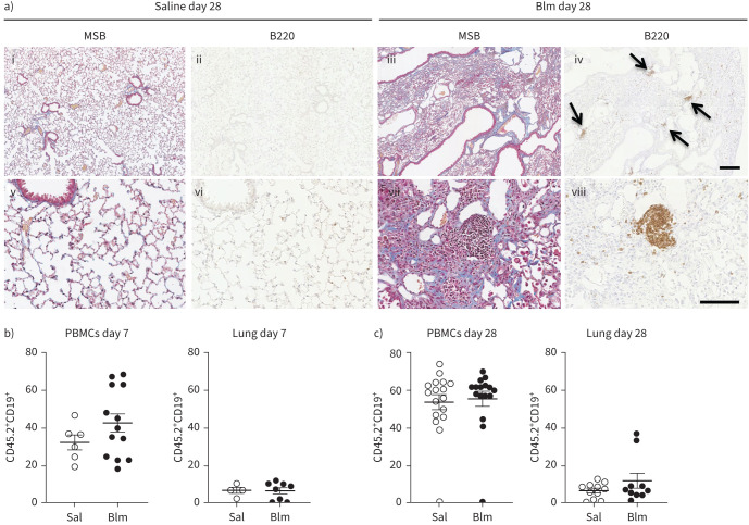
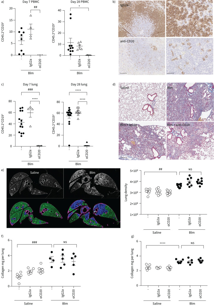
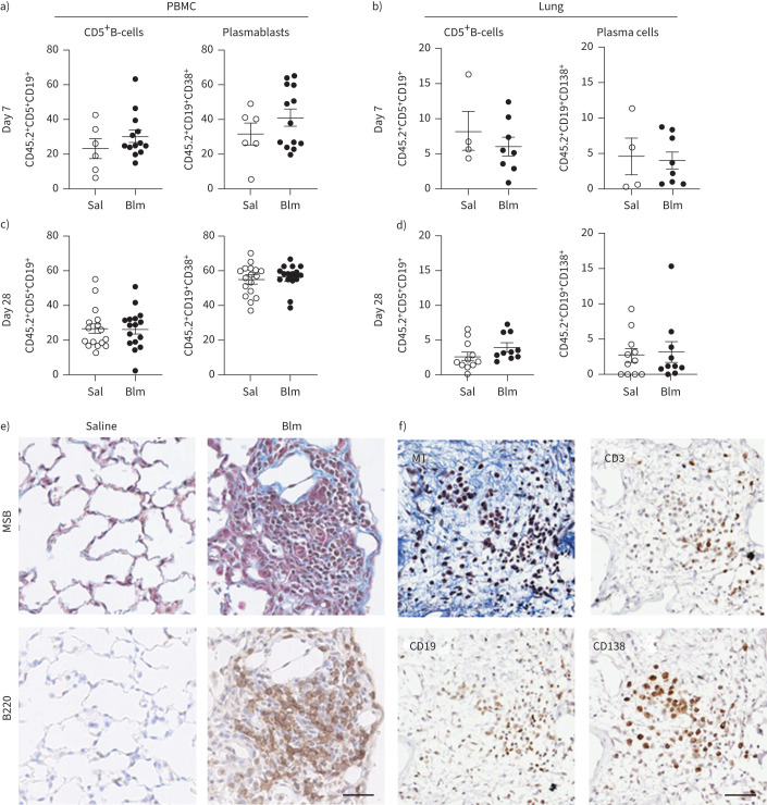
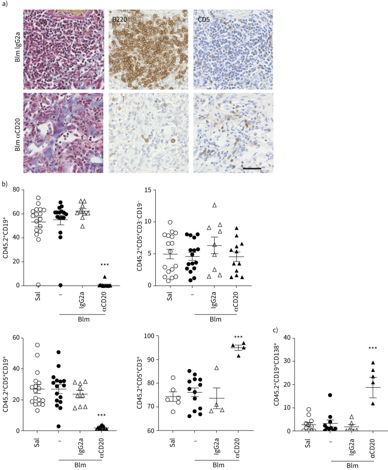
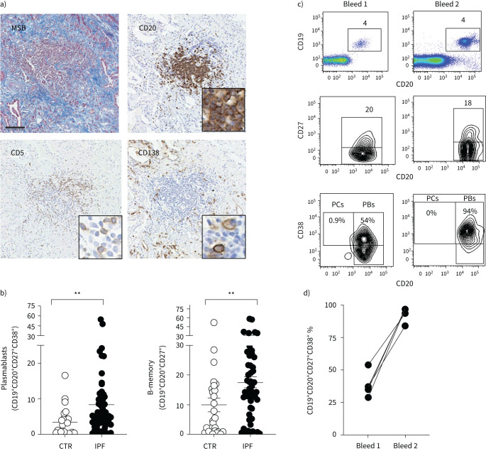
Idiopathic pulmonary fibrosis (IPF) is an interstitial lung disease associated with chronic inflammation and tissue remodelling leading to fibrosis, reduced pulmonary function, respiratory failure and death. Bleomycin (Blm)-induced lung fibrosis in mice replicates several clinical features of human IPF, including prominent lymphoid aggregates of predominantly B-cells that accumulate in the lung adjacent to areas of active fibrosis. We have shown previously a requirement for B-cells in the development of Blm-induced lung fibrosis in mice. To determine the therapeutic potential of inhibiting B-cell function in pulmonary fibrosis, we examined the effects of anti-CD20 B-cell ablation therapy to selectively remove mature B-cells from the immune system and inhibit Blm-induced lung fibrosis. Anti-CD20 B-cell ablation did not reduce fibrosis in this model; however, immune phenotyping of peripheral blood and lung resident cells revealed that anti-CD20-treated mice retained a high frequency of CD19+ CD138+ plasma cells. Interestingly, high levels of CD138+ cells were also identified in the lung tissue of patients with IPF, consistent with the mouse model. Treatment of mice with bortezomib, which depletes plasma cells, reduced the level of Blm-induced lung fibrosis, implicating plasma cells as important effector cells in the development and progression of pulmonary fibrosis.


Conflict of interest statement

Conflict of interest: S.E. Mutsaers, C.M. Prêle, R.J. McAnulty, G.J. Laurent, G. Hoyne, D.A. Knight, R.J. O'Donoghue and M. Ernst received grants from National Health and Medical Research Council (NHMRC), project grants ID APP1067511. R.J. McAnulty, S.E. Mutsaers, C.M. Prêle and D.R. Pearce received grants from British Lung Foundation, project grant number PPRG15-10. T. Miles reports that Genentech provided the anti-CD20 antibody, and received UWA Research Training Program Scholarship and the Lung Foundation Australia Bill van Nierop PhD Scholarship. All other authors have nothing to disclose.

Figures

FIGURE 1
FIGURE 1
B-cells are a prominent feature in bleomycin (Blm)-induced pulmonary fibrosis in mice. a) Immunohistochemical analysis of B220 staining 28 days post-oropharyngeal Blm treatment reveals an increase in B220+ cells (iv, arrows, and viii) at sites of fibrosis (Martius scarlet blue (MSB) staining, iii, vii collagen blue) compared to saline-treated controls (MSB i, v) and B220-stained (ii, vii). Images i–iv represent low magnification; scale bar=200 µm. Images v–iv represent high magnification; scale bar=100 µm. Images are representative of three mice in each group. b) Flow cytometry did not show a significant increase in circulating or lung resident CD45.2+ CD19+ B-cells in Blm-treated mice 7 days post-treatment (n≥4). c) No significant difference in circulating CD45.2+ CD19+ B-cells was detected in isolated peripheral blood mononuclear cells (PBMCs) 28 days following Blm treatment. There was no significant difference in the number of CD19+ cells detected in dissociated lung tissue post-Blm treatment. Statistical analysis was performed using an unpaired t-test (n≥10).
FIGURE 2
FIGURE 2
Anti-CD20-mediated B-cell depletion does not protect mice against oropharyngeal bleomycin (Blm)-induced pulmonary fibrosis. a) Anti-CD20 therapy 7 days prior to and 7 days following Blm successfully reduced circulating CD19+ cells as measured by flow cytometry in mice on day 7 and 28 post Blm treatment (n≥4). b) Immunohistochemical analysis of B220 expression on spleen tissue sections demonstrated a marked decrease in B220+ cell numbers in anti-CD20-treated mice compared to IgG2a-treated control mice. Scale bars=100 µm (25 µm higher power). c) Quantitative flow cytometry analysis showed that anti-CD20 treatment significantly reduced Blm-induced CD19+ cell numbers in the lung on day 7 and day 28 (n≥4). d) Masson's trichrome staining showed no overt difference in the extent of collagen deposition (blue) in mice treated with Blm and anti-CD20 compared to Blm only and mice treated with Blm and IgG2a. Scale bars=200 µm. e–g) Quantitative analysis of fibrosis and collagen deposition was performed using micro computed tomography (μCT) and high-performance liquid chromatography (HPLC) analysis of hydroxyproline, but there was no significant difference in mice treated with Blm and anti-CD20 compared with Blm and vehicle control. e) Represents a preventative model where anti-CD20 was administered 7 days prior to, and 7 days after Blm treatment. μCT analysis was performed ex vivo and the lung density was used as a representation of fibrosis. There was no difference in the amount of Blm-induced fibrosis in IgG2a or αCD20 treated mice (n≥7). f) HPLC analysis did not show any difference in the amount of Blm-induced lung collagen in in IgG2a- or αCD20-treated mice, confirming that αCD20 had no effect on lung fibrosis (n≥6). g) The data represent a therapeutic model where anti-CD20 was administered on days 10 and 19 post-Blm treatment. Similarly, there was no detectable change in the amount of collagen in the lungs of Blm and anti-CD20 treated mice compared to Blm alone or Blm- and IgG2a-treated mice (HPLC n≥5 and μCT n≥6). All images are representative of at least three mice per group. *: p≤0.05, ##: p≤0.005, ###: p≤0.0005, ****: p≤0.0001.
FIGURE 3
FIGURE 3
Characterisation of specific B-cell subsets in the peripheral circulation and lung of mice following oropharyngeal bleomycin (Blm)-induced pulmonary fibrosis. a) Blm treatment had no effect on the number of CD5+CD19+ B-cells or CD19+CD38+CD138+ plasma cells detected in peripheral blood mononuclear cells (PBMCs) on day 7 post-Blm treatment (n≥6) or b) CD5+CD19+ B-cells or CD19+CD138+ PCs in the lung 7 days post-Blm treatment (n≥4). c, d) No significant difference was observed in the frequency of CD19+CD138+ plasma cells in the circulation or the lung at day 28 post-Blm treatment (n≥6). No significant increase in CD5+CD19+ cells was observed in the lung 28 days post-Blm treatment (n≥6). Statistical analysis was performed using an unpaired t-test. e) Mouse lung tissue from saline-treated or Blm-treated mice (day 28) was stained with Martius scarlet blue (MSB; collagen blue) or with anti-B220 antibody. B220+ cell aggregates were detected in the lungs of Blm-treated mice (brown 3,3′-diaminobenzidine staining). f) Serial sections of Blm-treated lung stained with Masson's trichrome (MT) and immune markers. CD3+ lymphocytes, CD19+ B-cells and CD138+ plasma cells were detected at sites of fibrosis. All images are representative of at least three mice per group. Scale bars=50 µm.
FIGURE 4
FIGURE 4
B-cell composition of the lungs of mice following anti-CD20 and bleomycin (Blm) treatment. a) Mouse lung tissue from Blm-exposed mice treated with anti-CD20 or isotype control (day 28) was stained with Masson's trichrome (collagen blue). Immunohistochemistry shows a marked reduction in B220+ B-cells in 28-day treated mice and retention of CD5+ expressing cells compared with mice treated with Blm and IgG2a. All images are representative of at least three mice per group. Scale bars=50 µm. b) Flow cytometry confirms depletion of CD19+ cells in the circulation, but revealed no increase in circulating CD5+ B-cells, but a significant increase in CD5+ T-cells following anti-CD20 depletion (n≥4). c) However, anti-CD20 treatment did not deplete the plasma cell population in the lung 28 days following Blm (n≥5). ns: nonsignificant. Statistical analysis was performed using one-way ANOVA. ***: p≤0.001.
FIGURE 5
FIGURE 5
Plasma cell depletion reduces bleomycin (Blm)-induced lung fibrosis. Mice were given either a vehicle control or bortezomib (Btz) at day −7 and twice weekly until 28 days after intranasal Blm (2 mg·kg−1) treatment (day 0) and the volume of fibrosis was quantified by micro-computed tomography analysis. a) Blm induced significant lung fibrosis compared to saline, which was reduced following bortezomib treatment (n≥4). b) Immunofluorescence staining of lung tissue from Blm+vehicle or bortezomib-treated mice was performed to detect plasma cells within the lungs. The nuclei are stained blue, CD19+ cells are stained red and CD138+ cells are stained green. The plasma cells are positive for both CD19 and CD138. Some epithelial cell staining is seen in the bortezomib-treated group. The figure shows a representative image from each group (n≥4). Scale bars=60 μm. DAPI: 4′,6-diamidino-2-phenylindole. *: p<0.05. A lower-power image showing an extensive area of fibrosis is shown in supplementary figure S3.
FIGURE 6
FIGURE 6
Analysis of B-cell populations in the lung and peripheral blood of idiopathic pulmonary fibrosis (IPF) patients and aged matched healthy controls (CTR). a) Martius scarlet blue (MSB) stained and anti-CD20 stained sections of IPF lung tissue demonstrate collagen deposition (blue) and an accumulation of CD20+ B-cells within regions of fibrosis (brown). In addition, CD5+ and CD138+ plasma cells were abundant in IPF lung tissue. All images are representative of at least three mice per group. Scale bars=50 µm. b) Quantitation of the frequency of plasmablasts and memory B-cells in white blood cells of healthy controls (n=27) and IPF patients (n=51) as determined by flow cytometry. Each symbol represents the value from one individual. c) Representative dot plot and contour plots of CD19+ CD20+ B-cells from an IPF patient who was bled on two separate occasions 18 months apart. CD19+ versus CD20+ staining identifies mature B-cells (top), CD20+CD27+ cells identifies memory B-cells (middle) and CD20+ CD38+ identifies plasmablasts (PBs) (bottom), n=4. d) Frequency of CD19+CD20+ CD27+ CD38+ plasmablasts in the peripheral blood of four different individuals bled on two occasions 18 months apart. PCs: plasma cells.

Comment in

References

    1. Lederer DJ, Martinez FJ. Idiopathic pulmonary fibrosis. N Engl J Med 2018; 378: 1811–1823. doi:10.1056/NEJMra1705751 - DOI - PubMed
    1. Maher TM, Evans IC, Bottoms SE, et al. . Diminished prostaglandin E2 contributes to the apoptosis paradox in idiopathic pulmonary fibrosis. Am J Respir Crit Care Med 2010; 182: 73–82. doi:10.1164/rccm.200905-0674OC - DOI - PMC - PubMed
    1. Richeldi L, Collard HR, Jones MG. Idiopathic pulmonary fibrosis. Lancet 2017; 389: 1941–1952. doi:10.1016/S0140-6736(17)30866-8 - DOI - PubMed
    1. Heukels P, van Hulst JAC, van Nimwegen M, et al. . Enhanced Bruton's tyrosine kinase in B-cells and autoreactive IgA in patients with idiopathic pulmonary fibrosis. Respir Res 2019; 20: 232. doi:10.1186/s12931-019-1195-7 - DOI - PMC - PubMed
    1. Hoyne GF, Elliott H, Mutsaers SE, et al. . Idiopathic pulmonary fibrosis and a role for autoimmunity. Immunol Cell Biol 2017; 95: 577–583. doi:10.1038/icb.2017.22 - DOI - PubMed

Publication types