. 2022 Jul 7;12(1):11544.
doi: 10.1038/s41598-022-14514-1.

Identification of early biomarkers in saliva in genetically engineered mouse model C(3)1-TAg of breast cancer

Affiliations

Identification of early biomarkers in saliva in genetically engineered mouse model C(3)1-TAg of breast cancer

Isadora Fernandes Gilson Sena et al. Sci Rep. .

Abstract

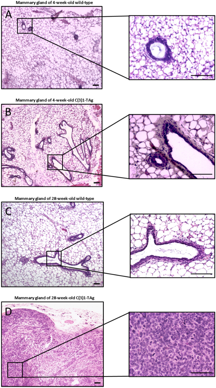
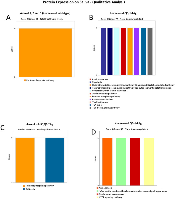
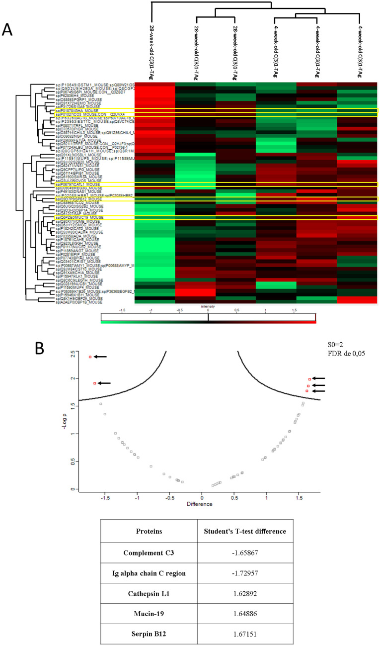
Breast cancer is one of leading causes of death worldwide in the female population. Deaths from breast cancer could be reduced significantly through earlier and more efficient detection of the disease. Saliva, an oral fluid that contains an abundance of protein biomarkers, has been recognized as a promising diagnostic biofluid that is easy to isolate through non-invasive techniques. Assays on saliva can be performed rapidly and are cost-effective. Therefore, our work aimed to identify salivary biomarkers present in the initial stages of breast cancer, where cell alterations are not yet detectable by histopathological analysis. Using state-of-the-art techniques, we employed a transgenic mouse model of mammary cancer to identify molecular changes in precancerous stage breast cancer through protein analysis in saliva. Through corroborative molecular approaches, we established that proteins related to metabolic changes, inflammatory process and cell matrix degradation are detected in saliva at the onset of tumor development. Our work demonstrated that salivary protein profiles can be used to identify cellular changes associated with precancerous stage breast cancer through non-invasive means even prior to biopsy-evident disease.


Conflict of interest statement

The authors declare no competing interests.

Figures

Figure 1
Figure 1
Characterization of carcinoma development in C(3)1-TAg and wild-type mice. (A) Mammary tissue from 4-week-old wild-type and (B) 4-week-old C(3)1-TAg mice (C) Mammary tissue from 28-week-old wild-type and (D) 28-week-old C(3)1-TAg. Low and high power magnifications are shown. Tissue from one representative animal of each group is shown. Scale bar: 50 μm.
Figure 2
Figure 2
Qualitative analysis of the main proteins pathways expressed in the saliva samples of 4-week-old wild-type mice (A) versus (BD) C3(1)-TAg mice. All three wild-type animals had the same protein pathway expression and are represented by Fig. 1A. Four-week-old C(3)1-TAg animals 1, 2 and 3 presented complex pathways and are represented separately (Fig. 1B–D).
Figure 3
Figure 3
Protein expression in saliva of 4-week-old C(3)1-TAg females compared to age-matched wild-type. (A) Heatmap of protein expression in saliva comparing 4-week-old C(3)1-TAg with 4-week-old wild-type animals. The two proteins that are significantly higher expressed on 4-week-old C(3)1-TAg animals compared to 4-week-old wild-type mice are highlight in yellow. (B) Volcano plot showing two proteins (represented by red dots with arrows. Each red dot represents a separate gene) that are significantly higher expressed on 4-week-old C(3)1-TAg animals compared to 4-week-old wild-type mice.
Figure 4
Figure 4
Qualitative pathway analysis of proteins expressed in the saliva of 28-week-old wild-type mice (A,B) and (CE) C3(1)-TAg mice. The graphs obtained by the Panther software show a singular profile of protein expression of each animal, with one wild-type mice that could not be classified in Panther software.
Figure 5
Figure 5
Differential protein expression in the saliva of C3(1)-TAg animals at 4-week and 28-week time points. (A) Heatmap comparing 4-week-old and 28-week-old C(3)1-TAg. The proteins that differ significantly between 4-week-old C(3)1-TAg animals and 4-week-old wild-type mice are highlight in yellow. (B) Volcano plot showing five proteins (represented by red dots with arrows. Each red dot represents a separate gene) that are expressed differently between animals with mammary cancer in the initial stage and animals with the invasive carcinoma. A negative result demonstrates that the protein is more expressed in the 28-week-old C(3)1-TAg samples and a positive result demonstrates that the protein is more expressed in the 4-weeks-old C(3)1-TAg.

References

    1. Sung H, et al. Global Cancer Statistics 2020: GLOBOCAN estimates of incidence and mortality worldwide for 36 cancers in 185 countries. CA Cancer J. Clin. 2021;71:209–249. doi: 10.3322/caac.21660. - DOI - PubMed
    1. Arellano M, et al. Current advances in identification of cancer biomarkers in saliva. Front. Biosci. (Schol. Ed.) 2009;1:296–303. doi: 10.2741/S27. - DOI - PubMed
    1. Harbeck N, et al. Breast cancer. Nat. Rev. Dis. Primers. 2019;5:66. doi: 10.1038/s41572-019-0111-2. - DOI - PubMed
    1. Drukteinis JS, Mooney BP, Flowers CI, Gatenby RA. Beyond mammography: New frontiers in breast cancer screening. Am. J. Med. 2013;126:472–479. doi: 10.1016/j.amjmed.2012.11.025. - DOI - PMC - PubMed
    1. Berg WA, et al. Diagnostic accuracy of mammography, clinical examination, US, and MR imaging in preoperative assessment of breast cancer. Radiology. 2004;233:830–849. doi: 10.1148/radiol.2333031484. - DOI - PubMed

Publication types