

Longitudinal study on background lesions in broiler breeder flocks and their progeny, and genomic characterisation of Escherichia coli
- PMID: 35799204
- PMCID: PMC9264609
- DOI: 10.1186/s13567-022-01064-7
Longitudinal study on background lesions in broiler breeder flocks and their progeny, and genomic characterisation of Escherichia coli
Abstract
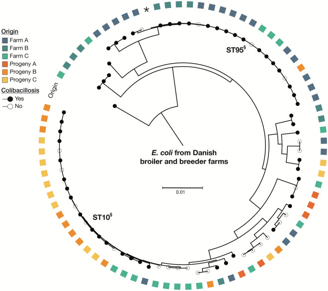
In broiler breeders, background mortality is rarely addressed, however, it represents the death of a vast number of birds, a constant productivity loss, welfare concerns and it might affect chick quality. The study aimed to unveil lesions leading to mortality in a study population perceived as healthy, combined with whole-genome sequencing (WGS) of Escherichia coli, a well-known contributor to disease problems in poultry. Broiler breeders (n = 340) originating from three distinct, putative healthy flocks and their progeny (n = 154) were subjected to a comprehensive post-mortem examination, bacteriological sampling, and sequencing of 77 E. coli isolates. Productivity data confirmed an exemplary health status of the enrolled flocks, and post-mortem examination further verified the absence of general disease problems. Among the submitted broiler breeders, exudative peritonitis (31.2%) was the most frequent lesion linked to infectious disease, whereas airsacculitis, pericarditis, perihepatitis, and salpingitis occurred in 18.5%, 3.5%, 3.8% and 17%, respectively. Yolksacculitis occurred in 15.6% of the broilers, whilst pericarditis, perihepatitis and peritonitis were diagnosed in 9.7%, 7.1% and 9.1%, respectively. WGS revealed a diverse population where ST95 dominated the population retrieved from broiler breeders, whereas ST10 was highly prevalent among broilers. Both lineages could be isolated from extraintestinal sites of birds without lesions indicative of infection. In general, the genetic diversity within flocks was comparable to the diversity between farms, and the overall occurrence of resistance markers was low. In conclusion, a comprehensive insight into lesions associated with background mortality is presented, together with a vast diversity of E. coli isolated from extraintestinal sites during a non-outbreak situation.
Keywords: APEC; Escherichia coli; Mortality; antimicrobial resistance; avian pathogenic E. coli; colibacillosis; pathology; surveillance; whole-genome sequencing.
© 2022. The Author(s).
Conflict of interest statement
The authors declare that they have no competing interests.
Figures

Similar articles
-
A molecular epidemiological study on Escherichia coli in young chicks with colibacillosis identified two possible outbreaks across farms.Vet Res. 2023 Feb 6;54(1):10. doi: 10.1186/s13567-023-01140-6. Vet Res. 2023. PMID: 36747303 Free PMC article.
-
Virulence of Escherichia coli Isolates Obtained from Layer Chickens with Colibacillosis Associated with Pericarditis, Perihepatitis, and Salpingitis in Experimentally Infected Chicks and Embryonated Eggs.Avian Dis. 2018 Jun;62(2):233-236. doi: 10.1637/11685-060717-ResNote.1. Avian Dis. 2018. PMID: 29944397
-
Dramatic increase in slaughter condemnations due to Escherichia coli ST23 and ST101 within the Danish broiler production.Vet Microbiol. 2023 May;280:109696. doi: 10.1016/j.vetmic.2023.109696. Epub 2023 Feb 19. Vet Microbiol. 2023. PMID: 36893553
-
[Escherichia coli salpingitis and peritonitis in layer chickens: an overview].Tijdschr Diergeneeskd. 2006 Nov 15;131(22):814-22. Tijdschr Diergeneeskd. 2006. PMID: 17263015 Review. Dutch.
-
[Avian pathogenic Escherichia coli (APEC)].Berl Munch Tierarztl Wochenschr. 2003 Sep-Oct;116(9-10):381-95. Berl Munch Tierarztl Wochenschr. 2003. PMID: 14526468 Review. German.
Cited by
-
A molecular epidemiological study on Escherichia coli in young chicks with colibacillosis identified two possible outbreaks across farms.Vet Res. 2023 Feb 6;54(1):10. doi: 10.1186/s13567-023-01140-6. Vet Res. 2023. PMID: 36747303 Free PMC article.
-
EspE3 plays a role in the pathogenicity of avian pathogenic Escherichia coli.Vet Res. 2023 Aug 29;54(1):70. doi: 10.1186/s13567-023-01202-9. Vet Res. 2023. PMID: 37644523 Free PMC article.
References
-
- Nolan LK, Vaillancourt J-P, Barbieri NL, Logue CM. Colibacillosis. In: Swayne DE, Boulianne M, Logue CM, McDougald LR, Nair V, Suarez DL, Wit S, Grimes T, Johnson D, Kromm M, Prajitno TY, Rubinoff I, Zavala G, editors. Diseases of Poultry. Hoboken: Wiley; 2020. pp. 770–830.
MeSH terms
Grants and funding
LinkOut - more resources
Full Text Sources
Medical
