

KAP1 phosphorylation promotes the survival of neural stem cells after ischemia/reperfusion by maintaining the stability of PCNA
- PMID: 35799276
- PMCID: PMC9264526
- DOI: 10.1186/s13287-022-02962-5
KAP1 phosphorylation promotes the survival of neural stem cells after ischemia/reperfusion by maintaining the stability of PCNA
Abstract
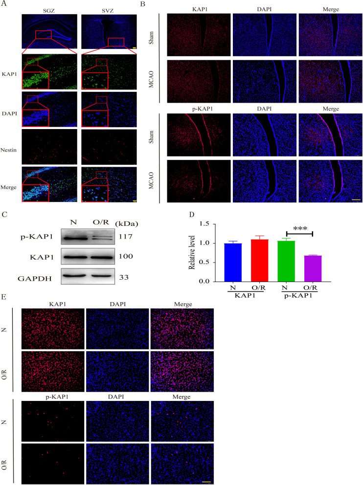
Aims: To explore the function of phosphorylation of KAP1 (p-KAP1) at the serine-824 site (S824) in the proliferation and apoptosis of endogenous neural stem cells (NSCs) after cerebral ischemic/reperfusion (I/R).
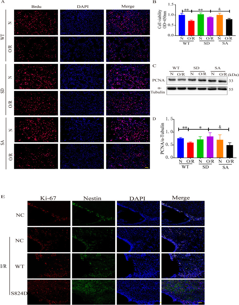
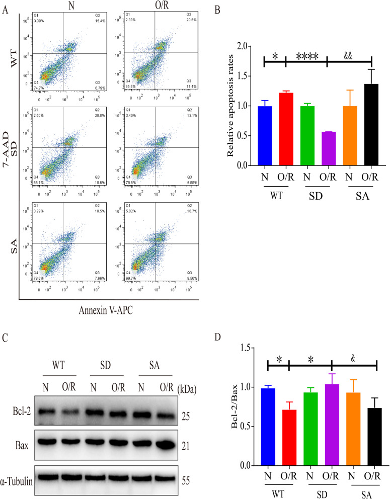
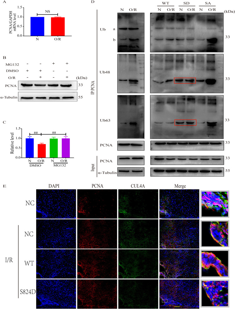
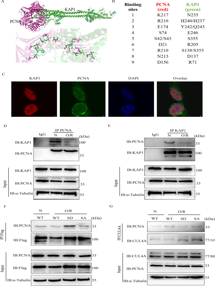
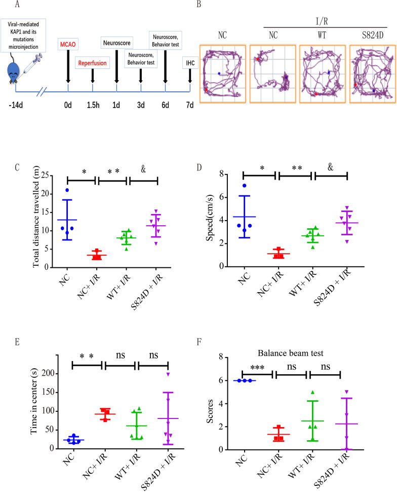
Methods: The apoptosis and proliferation of C17.2 cells transfected with the p-KAP1-expression plasmids and the expression of proliferation cell nuclear antigen (PCNA) and p-KAP1 were detected by immunofluorescence and Western blotting after the Oxygen Glucose deprivation/reperfusion model (OGD/R). The interaction of p-KAP1 and CUL4A with PCNA was analyzed by immunoprecipitation. In the rats MCAO model, we performed the adeno-associated virus (AAV) 2/9 gene delivery of p-KAP1 mutants to verify the proliferation of endogenous NSCs and the colocalization of PCNA and CUL4A by immunofluorescence.
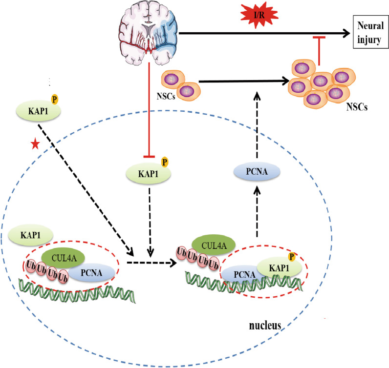
Results: The level of p-KAP1 was significantly down-regulated in the stroke model in vivo and in vitro. Simulated p-KAP1(S824) significantly increased the proliferation of C17.2 cells and the expression of PCNA after OGD/R. Simulated p-KAP1(S824) enhanced the binding of p-KAP1 and PCNA and decreased the interaction between PCNA and CUL4A in C17.2 cells subjected to OGD/R. The AAV2/9-mediated p-KAP1(S824) increased endogenous NSCs proliferation, PCNA expression, p-KAP1 binding to PCNA, and improved neurological function in the rat MCAO model.
Conclusions: Our findings confirmed that simulated p-KAP1(S824) improved the survival and proliferation of endogenous NSCs. The underlying mechanism is that highly expressed p-KAP1(S824) promotes binding to PCNA, and inhibits the binding of CUL4A to PCNA. This reduced CUL4A-mediated ubiquitination degradation to increase the stability of PCNA and promote the survival and proliferation of NSCs.
Keywords: Cerebral ischemia/reperfusion (I/R); KRAB domain protein 1(KAP1); Neural stem cells (NSCs); PCNA; Proliferation.
© 2022. The Author(s).
Conflict of interest statement
The authors declare that they have no competing interests.
Figures
Similar articles
-
Transplantation of Neural Stem Cells-Overexpressed Ku70 Improves Neurological Deficits in a Mice Model of Cerebral Ischemia Stroke.Neurochem Res. 2024 Mar;49(3):718-731. doi: 10.1007/s11064-023-04065-w. Epub 2023 Dec 8. Neurochem Res. 2024. PMID: 38063947
-
[Mechanism of tetramethylpyrazine combined with neural stem cells transplantation on cerebral ischemia-reperfusion rats].Zhongguo Zhong Yao Za Zhi. 2024 May;49(9):2316-2325. doi: 10.19540/j.cnki.cjcmm.20230821.401. Zhongguo Zhong Yao Za Zhi. 2024. PMID: 38812132 Chinese.
-
Panax notoginseng saponins stimulates the differentiation and neurite development of C17.2 neural stem cells against OGD/R injuries via mTOR signaling.Biomed Pharmacother. 2024 Mar;172:116260. doi: 10.1016/j.biopha.2024.116260. Epub 2024 Feb 21. Biomed Pharmacother. 2024. PMID: 38382327
-
Circular RNA cZNF292 silence alleviates OGD/R-induced injury through up-regulation of miR-22 in rat neural stem cells (NSCs).Artif Cells Nanomed Biotechnol. 2020 Dec;48(1):594-601. doi: 10.1080/21691401.2020.1725536. Artif Cells Nanomed Biotechnol. 2020. PMID: 32052645
-
Neuroprotection by mesenchymal stem cell (MSC) administration is enhanced by local cooling infusion (LCI) in ischemia.Brain Res. 2019 Dec 1;1724:146406. doi: 10.1016/j.brainres.2019.146406. Epub 2019 Aug 24. Brain Res. 2019. PMID: 31454517 Review.
Cited by
-
Melatonin improves stroke through MDM2-mediated ubiquitination of ACSL4.Aging (Albany NY). 2024 Jan 29;16(2):1925-1937. doi: 10.18632/aging.205469. Epub 2024 Jan 29. Aging (Albany NY). 2024. PMID: 38289595 Free PMC article.
-
Ginkgo biloba extract mediates HT22 cell proliferation and migration after oxygen-glucose deprivation/reoxygenation via regulating RhoA-ROCK2 signalling pathway.Metab Brain Dis. 2025 Jan 7;40(1):91. doi: 10.1007/s11011-024-01502-9. Metab Brain Dis. 2025. PMID: 39775993 Free PMC article.
-
LncRNA regulation in ischemic stroke and their application prospects.Neural Regen Res. 2026 Mar 1;21(3):1058-1073. doi: 10.4103/NRR.NRR-D-24-00924. Epub 2025 Mar 25. Neural Regen Res. 2026. PMID: 40145979 Free PMC article.
-
Gene therapy of adeno-associated virus (AAV) vectors in preclinical models of ischemic stroke.CNS Neurosci Ther. 2023 Dec;29(12):3725-3740. doi: 10.1111/cns.14392. Epub 2023 Aug 8. CNS Neurosci Ther. 2023. PMID: 37551863 Free PMC article. Review.
-
Saponins enhance the stability and cost-efficiency of human embryonic stem cell culture.Cell Regen. 2025 Jan 21;14(1):3. doi: 10.1186/s13619-024-00220-y. Cell Regen. 2025. PMID: 39836295 Free PMC article.
References
Publication types
MeSH terms
Substances
LinkOut - more resources
Full Text Sources
Molecular Biology Databases
Research Materials
Miscellaneous
