

Subcortical and hippocampal brain segmentation in 5-year-old children: Validation of FSL-FIRST and FreeSurfer against manual segmentation
- PMID: 35799402
- PMCID: PMC9543285
- DOI: 10.1111/ejn.15761
Subcortical and hippocampal brain segmentation in 5-year-old children: Validation of FSL-FIRST and FreeSurfer against manual segmentation
Abstract
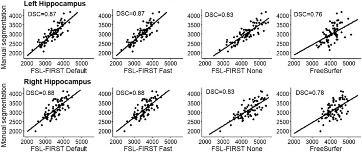
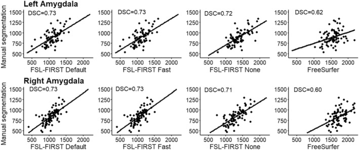
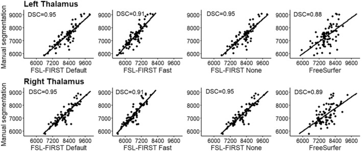
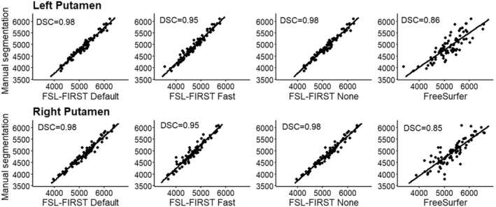
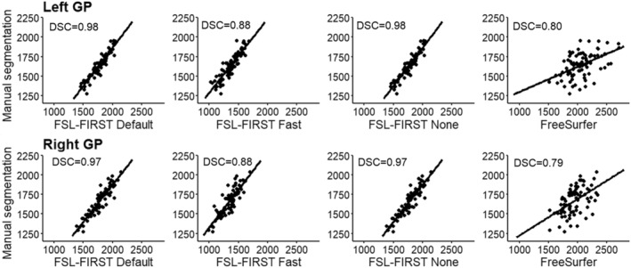
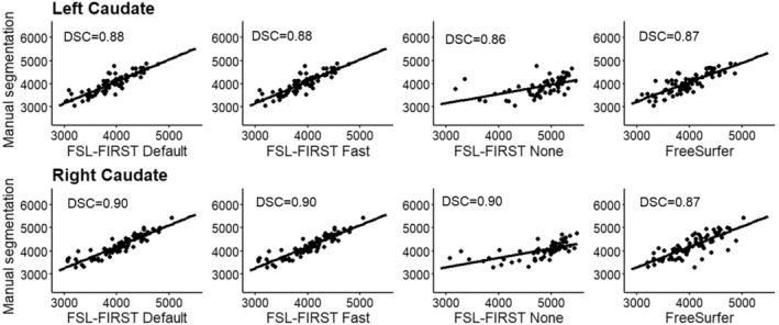
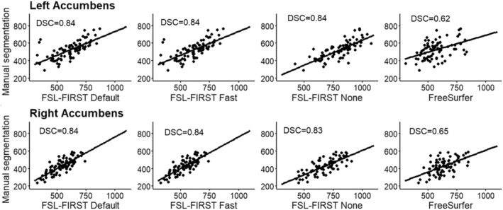
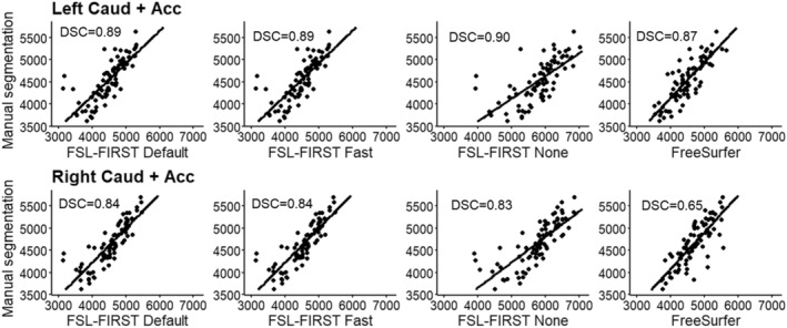
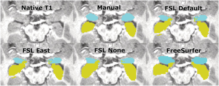
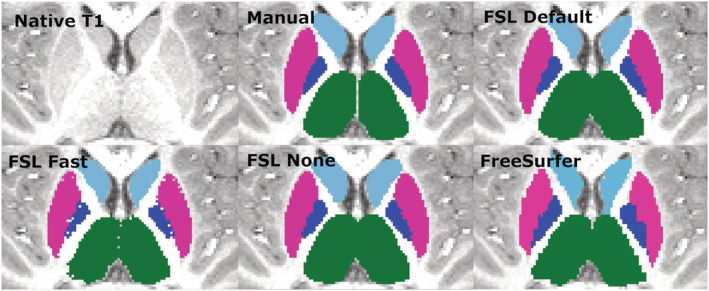
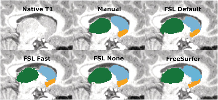
Developing accurate subcortical volumetric quantification tools is crucial for neurodevelopmental studies, as they could reduce the need for challenging and time-consuming manual segmentation. In this study, the accuracy of two automated segmentation tools, FSL-FIRST (with three different boundary correction settings) and FreeSurfer, were compared against manual segmentation of the hippocampus and subcortical nuclei, including the amygdala, thalamus, putamen, globus pallidus, caudate and nucleus accumbens, using volumetric and correlation analyses in 80 5-year-olds. Both FSL-FIRST and FreeSurfer overestimated the volume on all structures except the caudate, and the accuracy varied depending on the structure. Small structures such as the amygdala and nucleus accumbens, which are visually difficult to distinguish, produced significant overestimations and weaker correlations with all automated methods. Larger and more readily distinguishable structures such as the caudate and putamen produced notably lower overestimations and stronger correlations. Overall, the segmentations performed by FSL-FIRST's default pipeline were the most accurate, whereas FreeSurfer's results were weaker across the structures. In line with prior studies, the accuracy of automated segmentation tools was imperfect with respect to manually defined structures. However, apart from amygdala and nucleus accumbens, FSL-FIRST's agreement could be considered satisfactory (Pearson correlation > 0.74, intraclass correlation coefficient (ICC) > 0.68 and Dice score coefficient (DSC) > 0.87) with highest values for the striatal structures (putamen, globus pallidus, caudate) (Pearson correlation > 0.77, ICC > 0.87 and DSC > 0.88, respectively). Overall, automated segmentation tools do not always provide satisfactory results, and careful visual inspection of the automated segmentations is strongly advised.
Keywords: brain; brain (growth and development); child; neuroimaging.
© 2022 The Authors. European Journal of Neuroscience published by Federation of European Neuroscience Societies and John Wiley & Sons Ltd.
Conflict of interest statement
The authors declare no conflict of interest.
Figures
Similar articles
-
Hippocampus and amygdala volumes from magnetic resonance images in children: Assessing accuracy of FreeSurfer and FSL against manual segmentation.Neuroimage. 2016 Apr 1;129:1-14. doi: 10.1016/j.neuroimage.2016.01.038. Epub 2016 Jan 26. Neuroimage. 2016. PMID: 26824403 Free PMC article.
-
Evaluating accuracy of striatal, pallidal, and thalamic segmentation methods: Comparing automated approaches to manual delineation.Neuroimage. 2018 Apr 15;170:182-198. doi: 10.1016/j.neuroimage.2017.02.069. Epub 2017 Mar 1. Neuroimage. 2018. PMID: 28259781
-
A Comparative Analysis of MRI Automated Segmentation of Subcortical Brain Volumes in a Large Dataset of Elderly Subjects.Neuroinformatics. 2022 Jan;20(1):63-72. doi: 10.1007/s12021-021-09520-z. Epub 2021 Mar 30. Neuroinformatics. 2022. PMID: 33783668
-
A review on brain structures segmentation in magnetic resonance imaging.Artif Intell Med. 2016 Oct;73:45-69. doi: 10.1016/j.artmed.2016.09.001. Epub 2016 Sep 30. Artif Intell Med. 2016. PMID: 27926381 Review.
-
Looking beneath the surface: the importance of subcortical structures in frontotemporal dementia.Brain Commun. 2021 Jul 16;3(3):fcab158. doi: 10.1093/braincomms/fcab158. eCollection 2021. Brain Commun. 2021. PMID: 34458729 Free PMC article. Review.
Cited by
-
Prediction of East Asian Brain Age using Machine Learning Algorithms Trained With Community-based Healthy Brain MRI.Dement Neurocogn Disord. 2022 Oct;21(4):138-146. doi: 10.12779/dnd.2022.21.4.138. Epub 2022 Oct 31. Dement Neurocogn Disord. 2022. PMID: 36407289 Free PMC article.
-
Feasibility of FreeSurfer Processing for T1-Weighted Brain Images of 5-Year-Olds: Semiautomated Protocol of FinnBrain Neuroimaging Lab.Front Neurosci. 2022 May 2;16:874062. doi: 10.3389/fnins.2022.874062. eCollection 2022. Front Neurosci. 2022. PMID: 35585923 Free PMC article.
-
Deep learning segmentation of organs-at-risk with integration into clinical workflow for pediatric brain radiotherapy.J Appl Clin Med Phys. 2024 Mar;25(3):e14310. doi: 10.1002/acm2.14310. Epub 2024 Feb 19. J Appl Clin Med Phys. 2024. PMID: 38373283 Free PMC article.
-
A roadmap towards standardized neuroimaging approaches for human thalamic nuclei.Nat Rev Neurosci. 2024 Dec;25(12):792-808. doi: 10.1038/s41583-024-00867-1. Epub 2024 Oct 17. Nat Rev Neurosci. 2024. PMID: 39420114 Review.
-
Exploring the multidimensional nature of repetitive and restricted behaviors and interests (RRBI) in autism: neuroanatomical correlates and clinical implications.Mol Autism. 2023 Nov 27;14(1):45. doi: 10.1186/s13229-023-00576-z. Mol Autism. 2023. PMID: 38012709 Free PMC article.
References
-
- Acosta, H. , Kantojärvi, K. , Hashempour, N. , Pelto, J. , Scheinin, N. M. , Lehtola, S. J. , Lewis, J. D. , Fonov, V. S. , Collins, D. L. , Evans, A. , Parkkola, R. , Lähdesmäki, T. , Saunavaara, J. , Karlsson, L. , Merisaari, H. , Paunio, T. , Karlsson, H. , & Tuulari, J. J. (2020). Partial support for an interaction between a polygenic risk score for major depressive disorder and prenatal maternal depressive symptoms on infant right amygdalar volumes. Cerebral Cortex, 30, 6121–6134. 10.1093/cercor/bhaa158 - DOI - PubMed
-
- Akudjedu, T. N. , Nabulsi, L. , Makelyte, M. , Scanlon, C. , Hehir, S. , Casey, H. , Ambati, S. , Kenney, J. , O'Donoghue, S. , McDermott, E. , Kilmartin, L. , Dockery, P. , McDonald, C. , Hallahan, B. , & Cannon, D. M. (2018). A comparative study of segmentation techniques for the quantification of brain subcortical volume. Brain Imaging and Behavior, 12(6), 1678–1695. 10.1007/s11682-018-9835-y - DOI - PubMed
Publication types
MeSH terms
LinkOut - more resources
Full Text Sources
Medical
