. 2022 Jun 28:2022:8547563.
doi: 10.1155/2022/8547563. eCollection 2022.

CXCR4/CX43 Regulate Diabetic Neuropathic Pain via Intercellular Interactions between Activated Neurons and Dysfunctional Astrocytes during Late Phase of Diabetes in Rats and the Effects of Antioxidant N-Acetyl-L-Cysteine

Affiliations

CXCR4/CX43 Regulate Diabetic Neuropathic Pain via Intercellular Interactions between Activated Neurons and Dysfunctional Astrocytes during Late Phase of Diabetes in Rats and the Effects of Antioxidant N-Acetyl-L-Cysteine

Dan Zhu et al. Oxid Med Cell Longev. .

Abstract

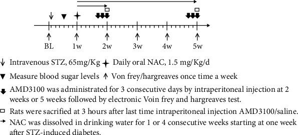
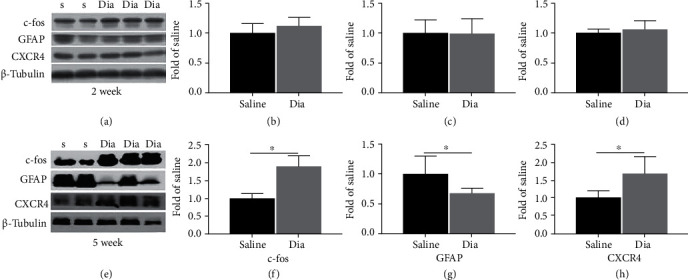
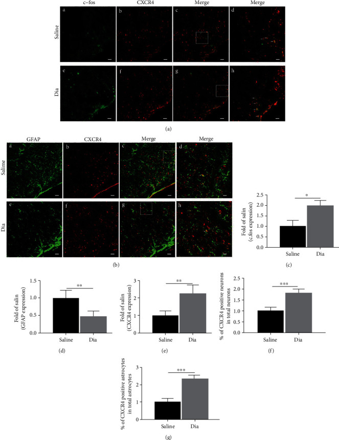
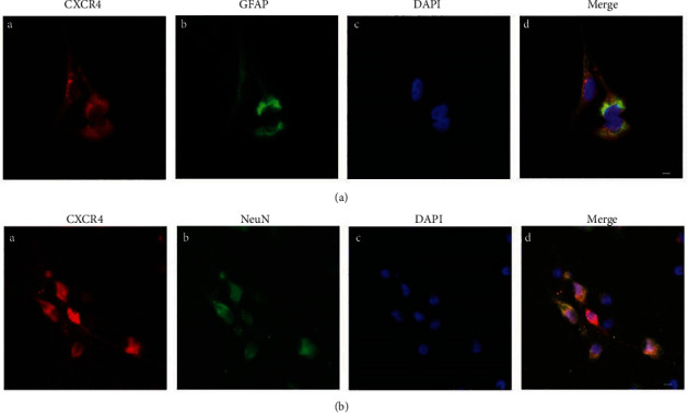
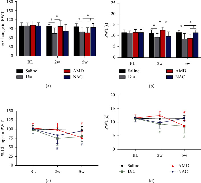
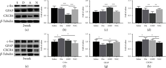
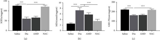
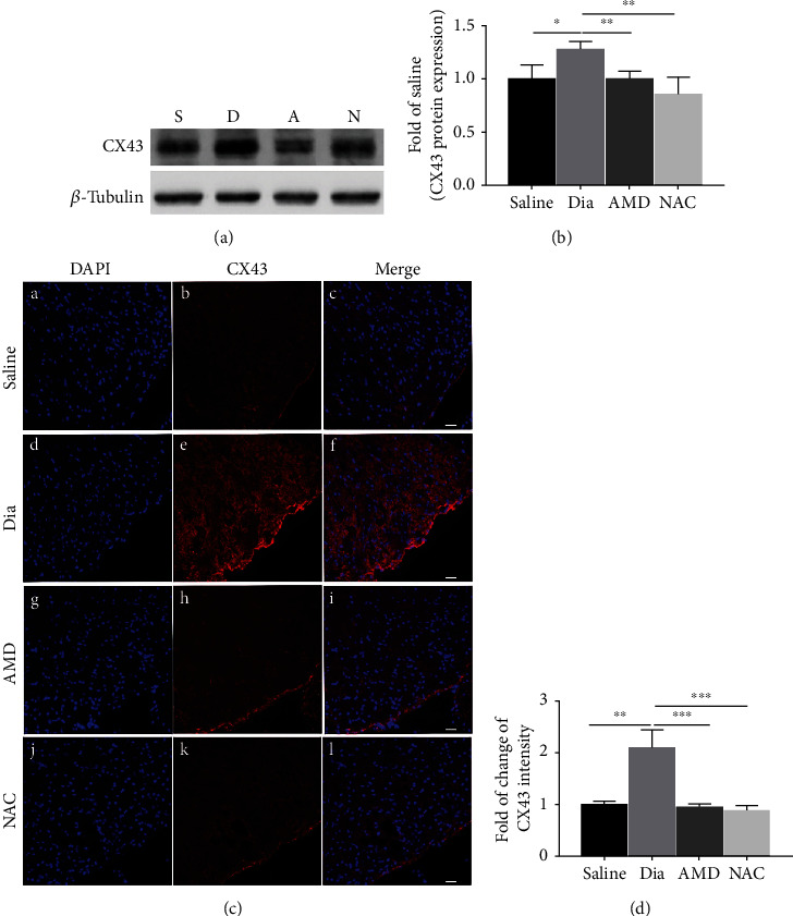
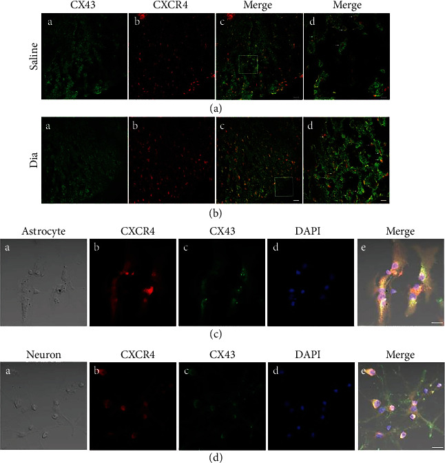
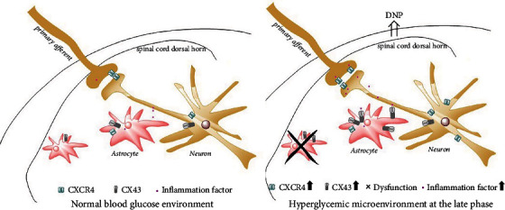
Growing evidence suggests that the interactions between astrocytes and neurons exert important functions in the central sensitization of the spinal cord dorsal horn in rodents with diabetes and neuropathic pain (DNP). However, it still remains unclear how signal transmission occurs in the spinal cord dorsal horn between astrocytes and neurons, especially in subjects with DNP. Chemokine CXC receptor 4 (CXCR4) plays critical roles in DNP, and connexin 43 (CX43), which is also primarily expressed by astrocytes, contributes to the development of neuropathy. We thus postulated that astrocytic and neuronal CXCR4 induces and produces inflammatory factors under persistent peripheral noxious stimulation in DNP, while intercellular CX43 can transmit inflammatory stimulation signals. The results showed that streptozotocin-induced type 1 diabetic rats developed heat hyperalgesia and mechanical allodynia. Diabetes led to persistent neuropathic pain. Diabetic rats developed peripheral sensitization at the early phase (2 weeks) and central sensitization at the late phase (5 weeks) after diabetes induction. Both CXCR4 and CX43, which are localized and coexpressed in neurons and astrocytes, were enhanced significantly in the dorsal horn of spinal cord in rats undergoing DNP during late phase of diabetes, and the CXCR4 antagonist AMD3100 reduced the expression of CX43. The nociceptive behavior was reversed, respectively, by AMD3100 at the early phase and by the antioxidant N-acetyl-L-cysteine (NAC) at the late phase. Furthermore, rats with DNP demonstrated downregulation of glial fibrillary acidic protein (GFAP) as well as upregulation of c-fos in the spinal cord dorsal horn at the late phase compared to the controls, and upregulation of GFAP and downregulation of c-fos were observed upon treatment with NAC. Given that GFAP and c-fos are, respectively, makers of astrocyte and neuronal activation, our findings suggest that CXCR4 as an inflammatory stimulation protein and CX43 as an intercellular signal transmission protein both may induce neurons excitability and astrocytes dysfunction in developing DNP.


Conflict of interest statement

This study is claimed to show no conflicts of interest.

Figures

Figure 1
Figure 1
Timeline of experiment protocol in SD rats. BL: baseline; w: week.
Figure 2
Figure 2
Streptozotocin (STZ)-induced DNP in rats enhanced spinal cord dorsal horn CXCR4 and c-fos and decreased GFAP at 5 weeks (e–h) yet not at 2 weeks (a–d) of diabetes. (a, e) c-fos, GFAP, and CXCR4 expression in the spinal cord, according to western blotting, at 2 and 5 weeks after intravenous STZ-induced diabetes (Dia) and at 2 and 5 weeks after injection of saline (saline control group). (b–d, f–h) Quantification of c-fos, GFAP, and CXCR4 levels in the spinal cord. Apart from that, western blot results can be shown to be means ± SD. ∗P < 0.05 in relative to saline group, n = 6/group. S = saline; Dia = diabetes.
Figure 3
Figure 3
Confocal images showed coexpression and distribution of CXCR4 and GFAP, CXCR4 and c-fos, and the expression of c-fos, CXCR4, and GFAP in the L3-L5 spinal cord dorsal horn in rats with STZ-induced DNP at 5 weeks of diabetes and in control rats receiving saline at 5 weeks. (a, b) Double-immunostaining for c-fos (green (a) (A and E)), GFAP (green (b) (A and E)), and CXCR4 (red, B and F) in the spinal dorsal horn after STZ-induced diabetes at 5 weeks. C and G were merged images of A and B or E and F separately (original magnification: 200×, scale bar A–C, E–G 20 μm). Merged and enlarged images were shown in D and H (original magnification: 400×, scale bar 10 μm). Immunostaining for c-fos (a) (A, E), GFAP (b) (A, E), and CXCR4 (B, F) of the spinal cord dorsal horn was determined at 5 weeks after STZ-induced diabetes. Besides, quantitative analysis of c-fos (c), GFAP (d), and CXCR4 (e) of the intensities at 5 weeks. Quantification of the coexpression of CXCR4 and c-fos (f) and CXCR4 and GFAP (g). All data are indicated to be means ± SD. ∗P < 0.05, ∗∗P < 0.01, and ∗∗∗P < 0.001 in relative to saline group. n = 6/group. .
Figure 4
Figure 4
Representative confocal images presented co-expression of CXCR4 and GFAP, CXCR4 and NeuN in primarily cultured astrocytes and neurons, respectively. (a, b) Double-immunostaining for CXCR4 (red, A), GFAP (astrocytic marker, green (a) (B)), NeuN (neuronal marker, green (b) (B)), DAPI (nuclear marker, blue, C). (a) (D) and (b) (D) were merged A, B, and C images (original magnification: 400×, scale bar 10 μm, n = 6/group).
Figure 5
Figure 5
Variations of PWT (a) and PWL (b) in untreated diabetic rats (Dia) and in nondiabetic control (saline) or diabetic rats exposed to treatment with AMD or NAC and variations of PWT (c) and PWL (d) from baseline in the saline, Dia, AMD, or NAC group. PWT (a) and PWL (b) were notably decreased in STZ-induced diabetic rats and increased in AMD-treated diabetic rats but not in NAC-treated diabetic rats at 2 weeks, while PWT and PWL were significantly increased in diabetic rats receiving oral NAC by 5 weeks as identified by electronic Von Frey and Hargreaves test. In addition, PWT (c) and PWL (d) were reduced from 2 weeks to 5 weeks compared to baseline in Dia group, and PWT (c) and PWL (d) were obviously lowered in 5 weeks in comparison with baseline in the AMD group. All findings are shown to be means ± SD, n = 6/group, ∗P < 0.05. #P < 0.05 vs. baseline. BL = baseline; w = week; PWT = paw withdraw threshold; PWL = paw withdrawal latencies; Dia = diabetes; NAC = N-acetyl-L-cysteine; AMD = AMD3100.
Figure 6
Figure 6
Correlative proinflammatory protein CXCR4 and neuronal marker protein c-fos of the spinal cord in rats with DNP was inhibited, and astrocytic marker protein GFAP was activated by intraperitoneal AMD3100 at 2 weeks and by daily oral NAC treatment at 5 weeks of diabetes. (a, e) Western blot demonstrated the protein expression of c-fos, GFAP, and CXCR4. (b–d, f–h) Quantitative analysis of c-fos, GFAP, and CXCR4 comparing with β-tubulin in spinal dorsal at 2 weeks and 5 weeks. In addition, all the obtained data can be shown to be means ± SD. ∗P < 0.05, n = 6/group.
Figure 7
Figure 7
Changes of antioxidant enzymes (SOD and GSH-PX) and lipid peroxidation product malondialdehyde (MDA) in the spinal cord in rats undergoing DNP at 5 weeks of diabetes and the treatment effects of NAC and AMD. (a–c) The activity of SOD (a) and the contents of MDA (b) and GSH-Px (c) in rat spinal cord at 5 weeks of diabetes. Besides, all the obtained data are denoted to be means ± SD. ∗P < 0.05, ∗∗P < 0.01, and ∗∗∗P < 0.001, n = 6/group.
Figure 8
Figure 8
DNP was associated with significantly added expression of CX43 in the spinal cord dorsal horn of STZ-induced diabetic rats at 5 weeks of the disease, which could be inhibited by intraperitoneal AMD3100 and NAC. (a) Western blot and (c) confocal images showed the protein expression of CX43 in the spinal cord dorsal horn. (b) Quantitative analysis of CX43 comparing with β-tubulin in the spinal dorsal at 5 weeks. All the data are demonstrated to be means ± SD. ∗P < 0.05, ∗∗P < 0.01, and ∗∗∗P < 0.001, n = 6/group. C, F, I, and M were merged images A and B, D and E, G and H, and J and K, separately. (d) Semiquantification of CX43 immunofluorescence intensity. n = 6/group (original magnification: 200×, scale bar 20 μm).
Figure 9
Figure 9
Confocal images show coexpression of CXCR4 and CX43 in the spinal dorsal horn of rats after STZ-induced DNP at 5 weeks of diabetes and in saline control rats (a, b), and in primary culture astrocytes (c) and neurons (d). (a) and (b) co-expression of CX43 and CXCR4 of spinal dorsal cord were shown in confocal images. (a) (A, B) and (b) (A, B) were double-immunostaining for CX43 (green, A) and CXCR4 (red, B). (a) (C) and (b) (C) were merged images A and B (original magnification: 200×, scale bar 20 μm). Merged and enlarged images were shown in (a) (D) and (b) (D) (original magnification: 400×, scale bar 10 μm, n = 6/group). (c) (A) and (d) (A) were bright field images of astrocytes and neurons, respectively. (c, d) Double-immunostaining for CXCR4 (red, B), CX43 (green, C), DAPI (blue, D). (c) (E) and (d) (E) were merged images A, B, C, and D (original magnification: 400×, scale bar 20 μm, n = 6/group).
Figure 10
Figure 10
Schematic hypothesis of neuronal-astrocytic CXCR4 and CX43-mediated diabetic neuropathic pain. STZ-induced type 1 diabetes resulted in persistent upregulation of CXCR4 and CX43 in astrocytes and neurons. It is shown that persistent increase of CXCR4 may cause activated neuron excitability and CX43 may mediate intercellular inflammation signal transmission, then dysfunctional astrocytes cannot counter the inflammatory factors at late phase of diabetes, and DNP occurs and exacerbates.

Similar articles

Cited by

References

    1. Feldman E. L., Nave K. A., Jensen T. S., Bennett D. L. H. New horizons in diabetic neuropathy: mechanisms, bioenergetics, and pain. Neuron . 2017;93(6):1296–1313. doi: 10.1016/j.neuron.2017.02.005. - DOI - PMC - PubMed
    1. Hur J., Sullivan K. A., Pande M., et al. The identification of gene expression profiles associated with progression of human diabetic neuropathy. Brain . 2011;134(11):3222–3235. doi: 10.1093/brain/awr228. - DOI - PMC - PubMed
    1. Thakur V., Sadanandan J., Chattopadhyay M. High-mobility group box 1 protein signaling in painful diabetic neuropathy. International Journal of Molecular Sciences . 2020;21(3):p. 881. doi: 10.3390/ijms21030881. - DOI - PMC - PubMed
    1. Wang Y., Li G., Stanco A., et al. CXCR4 and CXCR7 have distinct functions in regulating interneuron migration. Neuron . 2011;69(1):61–76. doi: 10.1016/j.neuron.2010.12.005. - DOI - PMC - PubMed
    1. Balabanian K., Lagane B., Infantino S., et al. The chemokine SDF-1/CXCL12 binds to and signals through the orphan receptor RDC1 in T lymphocytes. Journal of Biological Chemistry . 2005;280(42):35760–35766. doi: 10.1074/jbc.M508234200. - DOI - PubMed

MeSH terms