. 2022 Jun 5;12(11):e4431.
doi: 10.21769/BioProtoc.4431.

Protocol to Isolate Germinal Centers by Laser Microdissection

Affiliations

Protocol to Isolate Germinal Centers by Laser Microdissection

Farbod Bahreini et al. Bio Protoc. .

Abstract

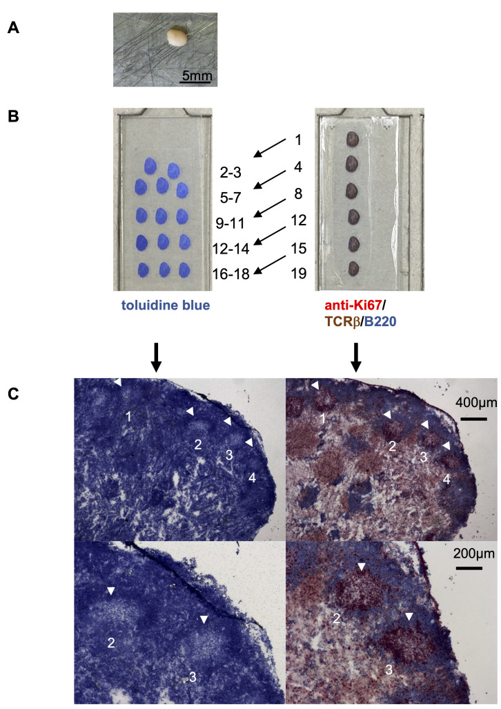
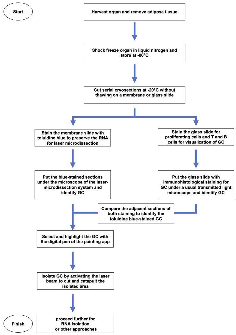
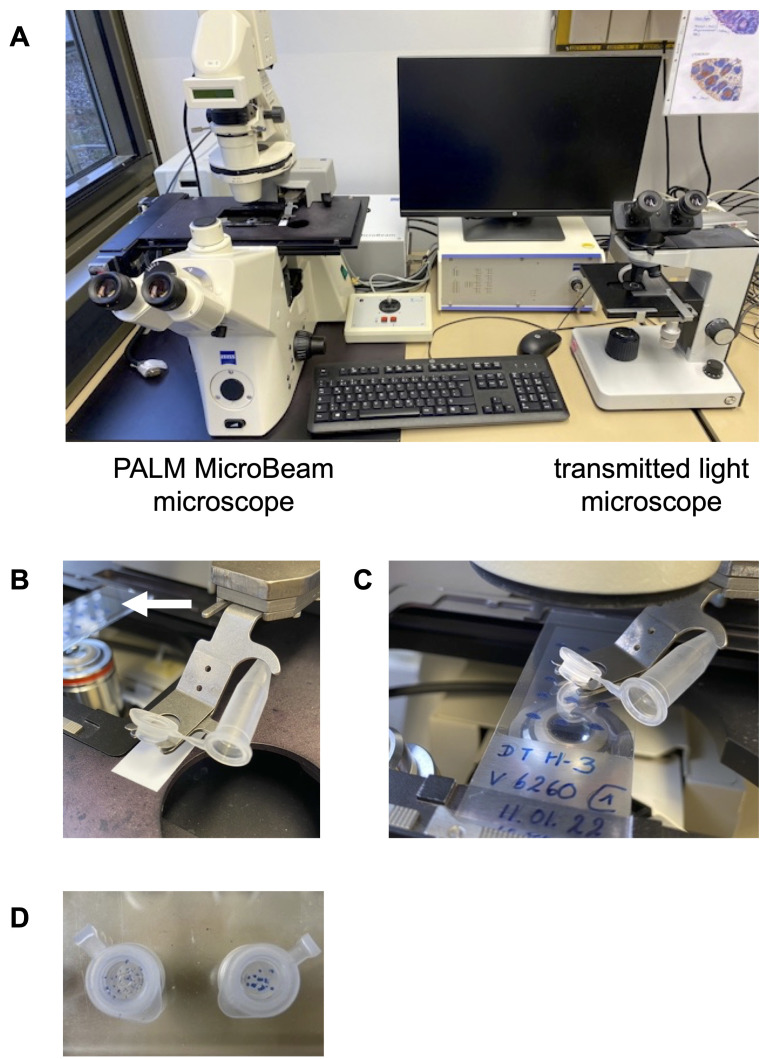
During adaptive immune responses, germinal centers (GC) appear as transient microstructures, in which antigen-specific B and T cells interact with each other. Because only the antigen-activated B and T cells, such as Plasmablasts or follicular T helper (Tfh) cells, are present in GC, the in depth-analysis of GC is of great interest. To identify the cells that reside within GC, the majority of studies use the expression of specific surface molecules for analysis by flow cytometry. To do so, the tissue has to be disrupted for the preparation of single-cell suspensions. Thereby, the local information regarding neighborhoods of B cells and T cells and their potential interaction is lost. To study GC in vivo within their original microenvironment, we established a protocol for the isolation of GC by laser microdissection. To enable the identification of GC for subsequent transcriptomic analysis, the degradation of mRNA was diminished by using frozen tissues and by establishing a rapid staining protocol. This procedure enables histological and transcriptomic analysis of individual GC even within one lymphoid organ.

Keywords: Cryo-preserved lymphoid structures; Follicular T helper cells; Germinal centers; In vivo analysis; Laser microdissection; Secondary lymphoid organs.


Conflict of interest statement

Competing interests The authors indicate no potential conflicts of interest.

Figures

Figure 1.
Figure 1.. Preparation of lymph nodes for isolation of GC by laser microdissection.
A. Activated murine popliteal lymph node. The surrounding adipose tissue was removed carefully before snap freezing. B. Serial cryosections of lymph nodes distributed on two slides, which were either stained with toluidine blue (left slide) or with anti-Ki67 (red) for proliferating cells, with anti-TCRβ for T cells (brown) and with anti-B220 for B cells (blue) (right side). The numbers indicate the order of serial sections. C. Example of two adjacent sections shown at higher magnifications. Toluidine blue-stained lymph node sections are shown on the left. Four GC are marked by white arrowheads (upper panel). Two GC are shown at a higher magnification on the lower panel. The immunohistochemical staining of the adjacent section on the right side confirms the identified GC (see Figure 2).
Figure 2.
Figure 2.. Workflow showing how the tissue is treated and processed in detail.
Video 1.
Video 1.. Laser capturing of lymphoid tissue compartments.
A toluidine blue-stained section of a rat spleen is used as an example for the microdissection of a B cell zone from lymphoid tissues. 1. The T- and B-cell zone can be clearly distinguished. 2. The area of interest, in this case, is a primary B cell follicle, which is circled using the drawing tool from the Palm Robo software. 3. The command “RoboLPC” from the Palm Robo software activates the laser beam to cut the marked tissue and the underlying membrane. An additional UV laser pulse catapults the isolated area into the cap of the microtube.
Figure 3.
Figure 3.. Equipment to isolate GC by laser microdissection.
A. The workplace shows the PALM MicroBeam microscope for laser capturing the tissues (left side) and the transmitted light microscope for immunohistochemical-stained sections to ensure the correct identification of GC (right side). B. A microtube is positioned into the holder. The white arrow indicates the membrane slide that contains the toluidine blue-stained sections in the slide holder. C. The cap- and microtube-holder must be placed directly over the slide to collect the catapulted tissues. D. The microdissected pieces of tissues can be clearly seen as toluidine blue-stained spots at the bottom of the microtube caps.

References

    1. Allen C. D. , Okada T. and Cyster J. G. ( 2007 . ). Germinal-center organization and cellular dynamics . Immunity 27 ( 2 ): 190 - 202 . - PMC - PubMed
    1. Cyster J. G. and Allen C. D. C. ( 2019 . ). B Cell Responses: Cell Interaction Dynamics and Decisions . Cell 177 ( 3 ): 524 - 540 . - PMC - PubMed
    1. Gasteiger G. , Ataide M. and Kastenmüller W. ( 2016 . ). Lymph node- an organ for T-cell activation and pathogen defense . Immunol Rev 271 ( 1 ): 200 - 220 . - PubMed
    1. Kalies K. , Konig P. , Zhang Y. M. , Deierling M. , Barthelmann J. , Stamm C. and Westermann J. ( 2008 . ). Nonoverlapping expression of IL10, IL12p40, and IFNgamma mRNA in the marginal zone and T cell zone of the spleen after antigenic stimulation . J Immunol 180 ( 8 ): 5457 - 5465 . - PubMed
    1. Niebuhr M. , Belde J. , Fahnrich A. , Serge A. , Irla M. , Ellebrecht C. T. , Hammers C. M. , Bieber K. , Westermann J. and Kalies K. ( 2021 . ). Receptor repertoires of murine follicular T helper cells reveal a high clonal overlap in separate lymph nodes in autoimmunity . Elife 10 : e70053 . - PMC - PubMed