Verbal Learning and Longitudinal Hippocampal Network Connectivity in Temporal Lobe Epilepsy Surgery
- PMID: 35800085
- PMCID: PMC9253296
- DOI: 10.3389/fneur.2022.854313
Verbal Learning and Longitudinal Hippocampal Network Connectivity in Temporal Lobe Epilepsy Surgery
Abstract
Introduction: Learning new verbal information can be impaired in 20-40% of patients after mesial temporal lobe resection. In recent years, understanding epilepsy as a brain network disease, and investigating the relationship between large-scale resting networks and cognition has led to several advances. Aligned studies suggest that it is the integrity of the hippocampal connectivity with these large-scale networks what is relevant for cognition, with evidence showing a functional and structural heterogeneity along the long axis hippocampus bilaterally.
Objective: Our aim is to examine whether pre-operative resting-state connectivity along the long hippocampal axis is associated with verbal learning decline after anterior temporal lobe resection.
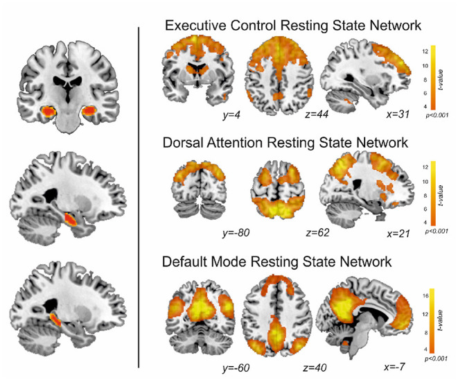
Methods: Thirty-one patients with epilepsy who underwent an anterior temporal lobe resection were pre-surgically scanned at 3-tesla, and pre/post-surgery evaluated for learning deficits using the Rey Auditory Verbal Learning Task (RAVLT). Eighteen controls matched by age, gender and handedness were also scanned and evaluated with the RAVLT. We studied the functional connectivity along the (anterior/posterior) long axis hippocampal subregions and resting-state functionally-defined brain networks involved in learning [executive (EXE), dorsal attention (DAN) and default-mode (DMN) networks]. Functional connectivity differences between the two groups of patients (learning intact or with learning decline) and controls were investigated with MANOVA and discriminant analysis.
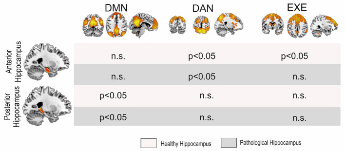
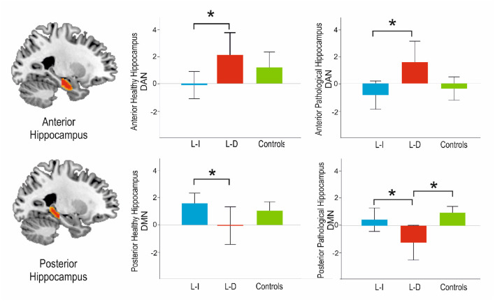
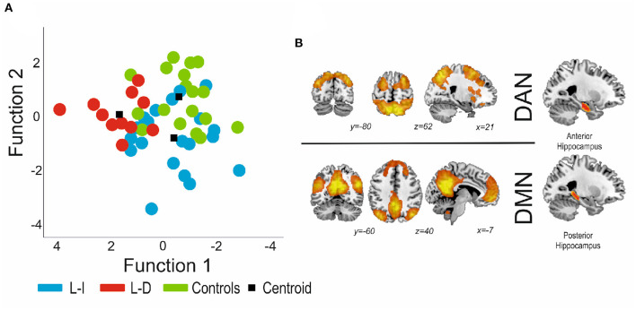
Results: There were significant differences in the pattern of hippocampal connectivity among the groups. Regarding the anterior connectivity hippocampal pattern, our data showed an increase of connectivity in the pathological side with the DAN (p = 0.011) and the EXE (p = 0.008) when comparing learning-decline vs. learning-intact patients. Moreover, the non-pathological side showed an increase in the anterior connectivity pattern with the DAN (p = 0.027) between learning-decline vs. learning-intact patients. In contrast, the posterior hippocampus showed a reduction of connectivity in the learning-decline patients with the DMN, both in the pathological (p = 0.004) and the non-pathological sides (p = 0.036). Finally, the discriminant analysis based on the pre-operative connectivity pattern significantly differentiated the learning-decline patients from the other groups (p = 0.019).
Conclusion: Our findings reveal bilateral connectivity disruptions along the longitudinal axis of the hippocampi with resting-state networks, which could be key to identify those patients at risk of verbal learning decline after epilepsy surgery.
Keywords: DMN (default mode network); dorsal attention network (DAN); resting-sate fMRI; temporal lobe epilepsy; verbal learning.
Copyright © 2022 Sala-Padro, Gifreu-Fraixino, Miró, Rodriguez-Fornells, Rico, Plans, Santurino, Falip and Càmara.
Conflict of interest statement
The authors declare that the research was conducted in the absence of any commercial or financial relationships that could be construed as a potential conflict of interest.
Figures