. 2022 Jun 24:20:472-488.
doi: 10.1016/j.bioactmat.2022.06.004. eCollection 2023 Feb.

Microenvironment-responsive electrocution of tumor and bacteria by implants modified with degenerate semiconductor film

Affiliations

Microenvironment-responsive electrocution of tumor and bacteria by implants modified with degenerate semiconductor film

Donghui Wang et al. Bioact Mater. .

Abstract

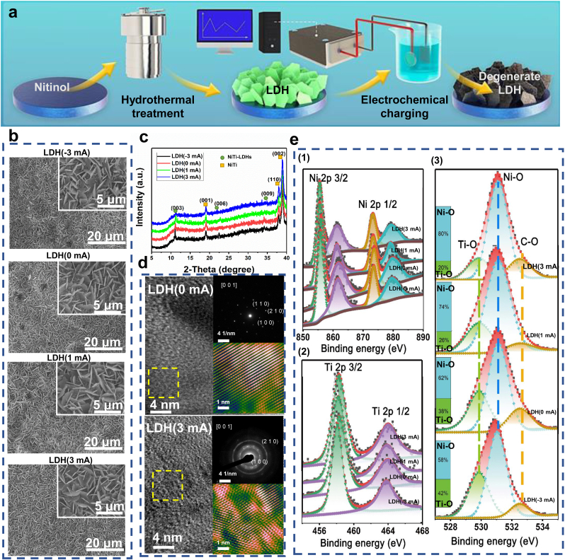
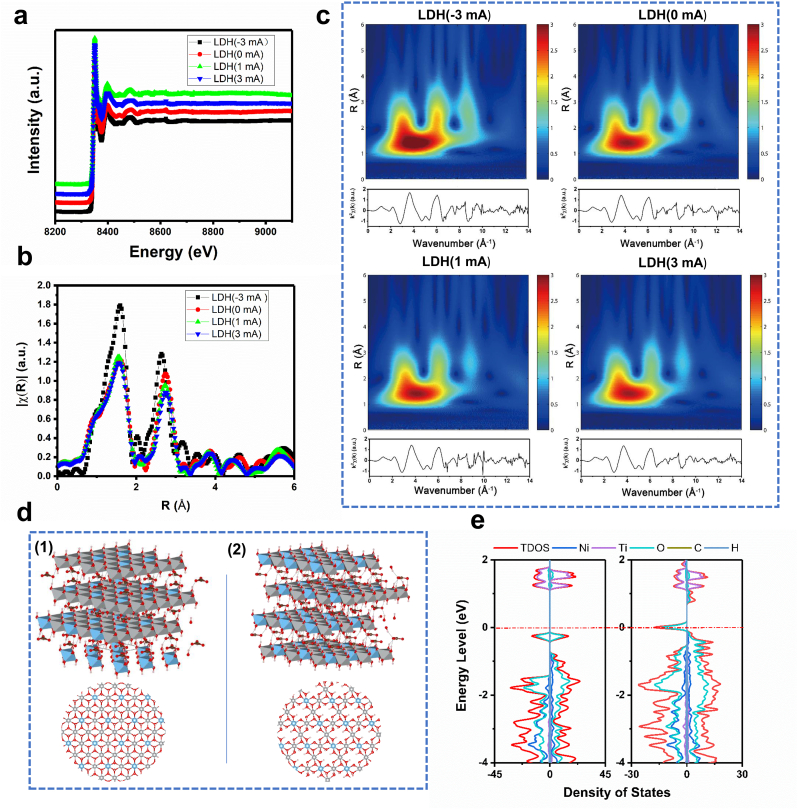
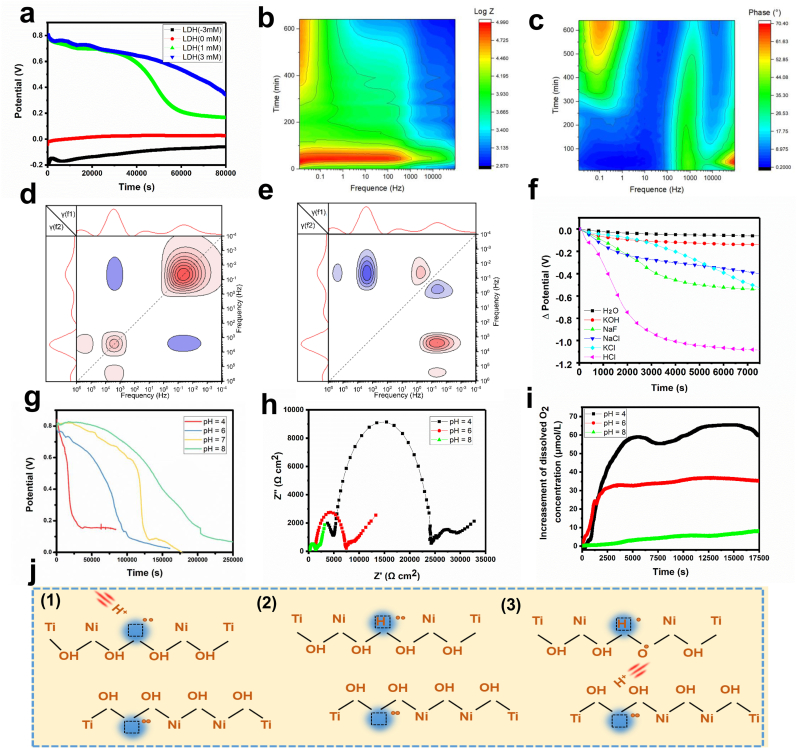
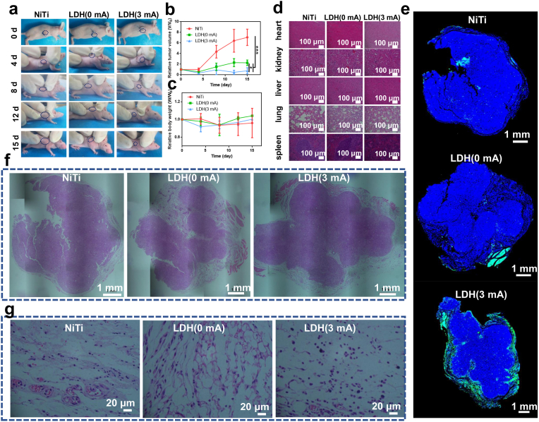
Implantable biomaterials are widely used in the curative resection and palliative treatment of various types of cancers. However, cancer residue around the implants usually leads to treatment failure with cancer reoccurrence. Postoperation chemotherapy and radiation therapy are widely applied to clear the residual cancer cells but induce serious side effects. It is urgent to develop advanced therapy to minimize systemic toxicity while maintaining efficient cancer-killing ability. Herein, we report a degenerate layered double hydroxide (LDH) film modified implant, which realizes microenvironment-responsive electrotherapy. The film can gradually transform into a nondegenerate state and release holes. When in contact with tumor cells or bacteria, the film quickly transforms into a nondegenerate state and releases holes at a high rate, rendering the "electrocution" of tumor cells and bacteria. However, when placed in normal tissue, the hole release rate of the film is much slower, thus, causing little harm to normal cells. Therefore, the constructed film can intelligently identify and meet the physiological requirements promptly. In addition, the transformation between degenerate and nondegenerate states of LDH films can be cycled by electrical charging, so their selective and dynamic physiological functions can be artificially adjusted according to demand.

Keywords: Anti-Tumor; Antibiosis; Electrotherapy; Implant; Layered double hydroxides.


Conflict of interest statement

The authors declare that they have no known competing financial interests or personal relationships that could have appeared to influence the work reported in this paper.

Figures

Image 1
Graphical abstract
Fig. 1
Fig. 1
Schematic illustration of H+-induced discharging of degenerate LDH films and their selective antitumor and antibacterial effects.
Fig. 2
Fig. 2
Preparation and characterization of degenerate LDH films on nitinol substrates. (a) Schematic diagram depicting the preparation process of degenerate LDH films. (b) SEM images of LDH(-3 mA), LDH(0 mA), LDH(1 mA) and LDH(3 mA) samples. (c) XRD patterns of LDH(-3 mA), LDH(0 mA), LDH(1 mA) and LDH(3 mA) samples. (d) HRTEM images of LDH(0 mA) and LDH(3 mA) samples. The insert image at the top right shows the SAED pattern, and the insert image at the bottom right shows the magnified image of the area marked by the yellow dotted box. (e) High-resolution XPS spectra of Ni (e−1), Ti (e−2), and O (e−3) acquired from LDH(-3 mA), LDH(0 mA), LDH(1 mA) and LDH(3 mA) samples.
Fig. 3
Fig. 3
Analysis and calculation of the coordination of Ni in degenerate LDH films. (a) Normalized XANES spectra at the Ni K-edge of LDH(-3 mA), LDH(0 mA), LDH(1 mA) and LDH(3 mA) samples. k2-weighted Fourier transform spectra from EXAFS spectra of LDH(-3 mA), LDH(0 mA), LDH(1 mA) and LDH(3 mA) samples. (c) Ni K-edge EXAFS spectra and modulus of their Morlet wavelet transform for LDH(-3 mA), LDH(0 mA), LDH(1 mA) and LDH(3 mA) samples. (d) Supercell models for chemometric NiTi LDHs (d-1) and NiTi LDHs with Ni vacancies (d-2), in which the gray ball represents Ni, the blue ball represents Ti, the red ball represents O, the pink ball represents H, and the brown ball represents C. (e) Total density of states of chemometric LDHs and LDHs with Ni vacancies.
Fig. 4
Fig. 4
Characterization of the conductivity and charge carriers in degenerate LDH films. (a) Photos showing the brightness of a small bulb in a constant voltage circuit (a-1) connected to LDH(-3 mA) (a-2), LDH(0 mA) (a-3), LDH(1 mA) (a-4) and LDH(3 mA) (a-5) samples. (b) I–V curves of LDH(-3 mA), LDH(0 mA), LDH(1 mA) and LDH(3 mA) samples measured in the dark. The inset image shows the test circuit. (c) I–V curves of LDH(-3 mA), LDH(0 mA), LDH(1 mA) and LDH(3 mA) samples. The curves were obtained under light, and the y-coordinate was plotted in logarithmic form. (d) MS curves of LDH(-3 mA), LDH(0 mA), LDH(1 mA) and LDH(3 mA) samples. (e) SECM images of LDH(-3 mA), LDH(0 mA), LDH(1 mA) and LDH(3 mA) samples when the applied voltage was 0.5 V. (f) SECM images of LDH(0 mA), LDH(1 mA) and LDH(3 mA) samples when the applied voltage was −0.5 V. (g) PACs of LDH(3 mA) samples with a positive or negative applied voltage. (h) Illustration of the redox transformation between ferrocene and ferrocenium in the electrolyte in SECM characterization. (i) Schematic diagram depicting the distribution of ferrocene/ferrocenium in the electrolyte when the voltage applied on the probe is positive or negative.
Fig. 5
Fig. 5
Hole transfer in the degenerate LDH films. (a) Polarization curves of LDH(-3 mA), LDH(0 mA), LDH(1 mA) and LDH(3 mA) samples. EIS Nyquist plots of LDH(-3 mA), LDH(0 mA), LDH(1 mA) and LDH(3 mA) samples. The inset image shows a magnified image of the area marked by the blue dotted box. (c) Impedance of LDH(-3 mA), LDH(0 mA), LDH(1 mA) and LDH(3 mA) samples acquired by EIS measurements. (d) Phase angle of LDH(-3 mA), LDH(0 mA), LDH(1 mA) and LDH(3 mA) samples acquired by EIS measurements. (e) Equivalent electrical circuits used for the fitting of EIS data of the LDH films in the nondegenerate state (e−1) and degenerate state (e−2). (f) DRT spectra of LDH(-3 mA), LDH(0 mA), LDH(1 mA) and LDH(3 mA) samples. (g) Water contact angles of LDH(-3 mA), LDH(0 mA), LDH(1 mA) and LDH(3 mA) samples.
Fig. 6
Fig. 6
H+-responsive hole release and transformation from the degenerate state to the nondegenerate state of degenerate LDH films. (a) OCP curves of LDH(-3 mA), LDH(0 mA), LDH(1 mA) and LDH(3 mA) samples. (b) Evolution of the impedance of the LDH(3 mA) sample acquired by sequential EIS measurements. (c) Evolution of the phase angle of the LDH(3 mA) sample acquired by sequential EIS measurements. (d) Two-dimensional DRT synchronous correlation spectrum of the LDH(3 mA) sample. (e) Two-dimensional DRT asynchronous correlation spectrum of the LDH(3 mA) sample. (f) OCP alteration of LDH(3 mA) samples immersed in electrolytes containing different types of ions. (g) OCP alteration of LDH(3 mA) samples immersed in electrolytes with different pH values. (h) EIS Nyquist plots of LDH(3 mA) immersed in electrolytes with different pH values. (i) Amounts of dissolved O2 in the solutions with different pH values in which LDH(3 mA) samples were immersed. (j) Diagram showing the mechanism resulting in H+-responsive hole release from degenerate LDH films.
Fig. 7
Fig. 7
In vitro selective cancer cell inhibition ability of degenerate LDH films. (a) Viability of cancer cells: proliferation of cancer cells cultured on various samples, n = 4 (a-1); live/dead stained cancer cells cultured on various samples (a-2); SEM images of cancer cells cultured on various samples, in which cells are marked by yellow (live), green (apoptotic), red (necrotic) and blue (early apoptotic) based on their survival state (a-3); percentages of cancer cells in different states acquired from the SEM images (a-4). (b) Viability of normal cells: proliferation of normal cells cultured on various samples, n = 4 (b-1); live/dead stained normal cells cultured on various samples (b-2); SEM images of normal cells cultured on various samples, in which cells were marked by yellow (live), green (apoptotic), red (necrotic) and blue (early apoptotic) based on their survival state (b-3); percentages of normal cells in different states acquired from the SEM images (b-4). (c) Expression of apoptosis-related genes in cancer cells and normal cells cultured on various samples: relative mRNA expression of Caspase-3 (c-1) and Bcl-2 (c-2) and ratio of Caspase-3/Bcl-2 (c-3) in cancer cells cultured on various samples; relative mRNA expression of Caspase-3 (c-4) and Bcl-2 (c-5) and ratio of Caspase-3/Bcl-2 (c-6) in normal cells cultured on various samples, n = 3.
Fig. 8
Fig. 8
In vivo tumor-inhibition effect of degenerate LDH films. (a) Representative photos of tumor-bearing mice after being implanted with NiTi, LDH(0 mA) and LDH(3 mA) samples for various time periods. (b) Relative tumor volumes of mice implanted with NiTi, LDH(0 mA) and LDH(3 mA) samples, n = 4. (c) Relative body weights of mice after being implanted with NiTi, LDH(0 mA) and LDH(3 mA) samples for various time periods, n = 4. (d) Images of H&E-stained spleen, lung, liver, kidney and heart tissue in mice implanted with NiTi, LDH(0 mA) and LDH(3 mA) samples. (e) Images of TUNEL-stained tumor tissue in mice implanted with NiTi, LDH(0 mA) and LDH(3 mA) samples. (f) Images of H&E-stained tumor tissue in mice implanted with NiTi, LDH(0 mA) and LDH(3 mA) samples. (g) Magnified images showing the periphery of the tumor tissue in mice implanted with NiTi, LDH(0 mA) and LDH(3 mA) samples.
Fig. 9
Fig. 9
Cyclic transformation of LDH films between nondegenerate and degenerate states. (a) OCP and water contact angle of LDH films after cyclic charging and discharging treatments, n = 3. (b) Photos of recultivated E. coli dissociated from various samples and corresponding colony number, n = 3. (c) Live/dead stained E. coli cultured on various samples. (d) SEM images of E. coli cultured on various samples. (e) Scheme illustrating the possible application of degenerate LDH films in the surface modification of various implants, realizing dynamic and selective regulation of the biological behavior in a long-lasting manner by coupling with wired and wireless power supplies.

References

    1. Dagenais G.R., Leong D.P., Rangarajan S., Lanas F., Lopez-Jaramillo P., Gupta R., Diaz R., Avezum A., Oliveira G.B.F., Wielgosz A., Parambath S.R., Mony P., Alhabib K.F., Temizhan A., Ismail N., Chifamba J., Yeates K., Khatib R., Rahman O., Zatonska K., Kazmi K., Wei L., Zhu J., Rosengren A., Vijayakumar K., Kaur M., Mohan V., Yusufali A., Kelishadi R., Teo K.K., Joseph P., Yusuf S. Variations in common diseases, hospital admissions, and deaths in middle-aged adults in 21 countries from five continents (PURE): a prospective cohort study. Lancet. 2020;395(10226):785–794. - PubMed
    1. Tchelebi L.T., Eng C., Messick C.A., Hong T.S., Ludmir E.B., Kachnic L.A., Zaorsky N.G. Current treatment and future directions in the management of anal cancer, CA-Cancer. J. Clin. 2021 - PubMed
    1. Zhu C., He M., Sun D., Huang Y., Huang L., Du M., Wang J., Wang J., Li Z., Hu B., Song Y., Li Y., Feng G., Liu L., Zhang L. 3D-Printed multifunctional polyetheretherketone bone scaffold for multimodal treatment of osteosarcoma and osteomyelitis. ACS Appl. Mater. Interfaces. 2021;13(40):47327–47340. - PubMed
    1. Bastiancich C., Malfanti A., Preat V., Rahman R. Rationally designed drug delivery systems for the local treatment of resected glioblastoma. Adv. Drug Deliv. Rev. 2021;177 - PubMed
    1. Gauto E., Fratti J.D.C., Salazar M., Macchi H., Banskota S.U.U., Baral B., Weir C., Ahuja K., Cattoni J.A. In-hospital outcomes of percutaneous coronary intervention with drug-eluting stent in patients with localized and metastatic cancer. J. Clin. Oncol. 2021;39(15)

LinkOut - more resources