

A case report of differentiated thyroid cancer presenting as a renal mass
- PMID: 35800419
- PMCID: PMC9205841
- DOI: 10.22038/AOJNMB.2021.60302.1422
A case report of differentiated thyroid cancer presenting as a renal mass
Abstract
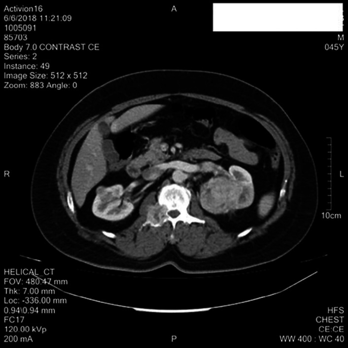
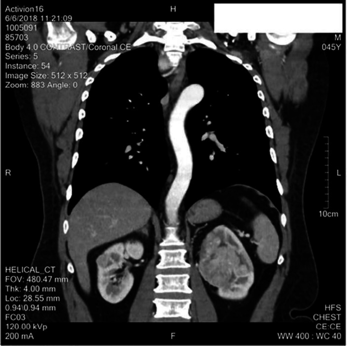
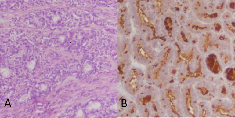
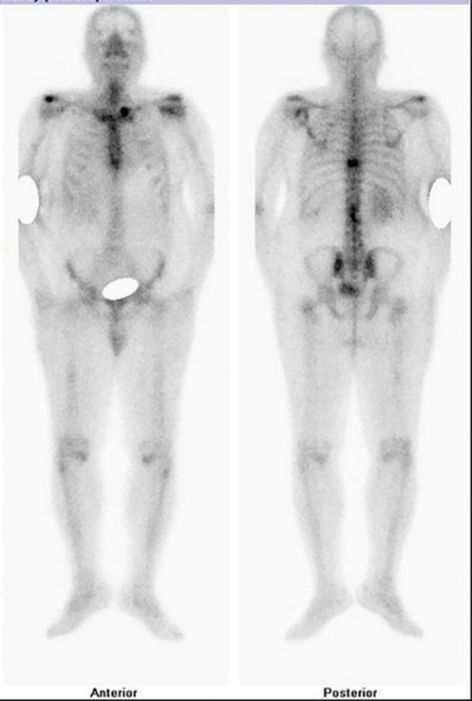
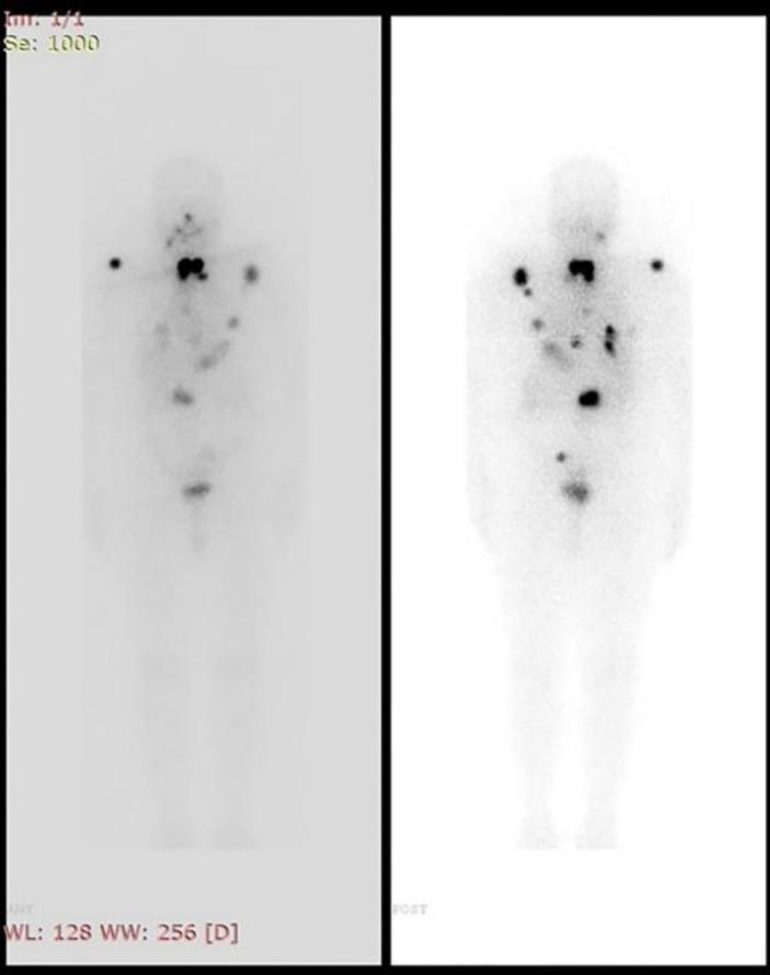
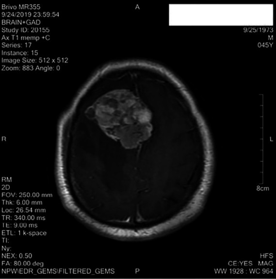
The kidney is an unconventional site for thyroid metastasis. As of the writing of this article, only about 30 cases have been reported. It presents like a renal mass. We are reporting a man with thyroid carcinoma presenting with distant metastasis to the kidney. He had complaints of abdominal pain and haematuria. Initial imaging suggested a left renal mass. A diagnosis of renal cell carcinoma was made and a nephrectomy was performed. Histopathology revealed it to be a metastasis from cancer of the thyroid gland. Subsequently, an ultrasound of the thyroid gland was performed, which showed a malignant appearing thyroid nodule. Correlative bone scan showed uptake at multiple skeletal sites. Total thyroidectomy was done and it was found to be papillary thyroid cancer. Subsequently, high dose radioactive iodine was administered. The patient was followed up and has recently found to have metastasis to the brain and is undergoing radiotherapy.
Keywords: Differentiated thyroid cancer; Papillary thyroid cancer; Renal metastasis.
© 2022 mums.ac.ir All rights reserved.
Figures
Similar articles
-
Papillary thyroid carcinoma presenting as a primary renal tumor with multiple pulmonary and bone metastases: a case report.J Med Case Rep. 2019 Apr 20;13(1):95. doi: 10.1186/s13256-019-2025-8. J Med Case Rep. 2019. PMID: 31003598 Free PMC article.
-
Renal Metastasis and Dual (18F-Fluorodeoxyglucose and 131I) Avid Skeletal Metastasis in a Patient with Papillary Thyroid Cancer.Indian J Nucl Med. 2017 Jan-Mar;32(1):50-53. doi: 10.4103/0972-3919.198482. Indian J Nucl Med. 2017. PMID: 28242987 Free PMC article.
-
Distant, solitary skeletal muscle metastasis in recurrent papillary thyroid carcinoma.Thyroid. 2011 Sep;21(9):1027-31. doi: 10.1089/thy.2010.0249. Epub 2011 Aug 11. Thyroid. 2011. PMID: 21834676
-
[Clinicopathologic characteristics of thyroid-like follicular carcinoma of the kidney: an analysis of five cases and review of literature].Zhonghua Bing Li Xue Za Zhi. 2016 Oct 8;45(10):687-691. doi: 10.3760/cma.j.issn.0529-5807.2016.10.004. Zhonghua Bing Li Xue Za Zhi. 2016. PMID: 27760609 Review. Chinese.
-
Distant metastases from thyroid and parathyroid cancer.ORL J Otorhinolaryngol Relat Spec. 2001 Jul-Aug;63(4):243-9. doi: 10.1159/000055749. ORL J Otorhinolaryngol Relat Spec. 2001. PMID: 11408821 Review.
References
-
- Li M, Dal Maso L, Vaccarella S. Global trends in thyroid cancer incidence and the impact of overdiagnosis. The Lancet Diabetes & Endocrinology. 2020;8(6):468–70. - PubMed
-
- Perros P, Boelaert K, Colley S, Evans C, Evans RM, Gerrard Ba G, et al. Guidelines for the management of thyroid cancer. Clinical endocrinology. 2014;81:1–122. - PubMed
Publication types
LinkOut - more resources
Full Text Sources
Research Materials