

Review
doi: 10.4103/jfmpc.jfmpc_1479_21.
Epub 2022 May 14.
Laboratory diagnosis of mucormycosis: Present perspective
Affiliations
- PMID: 35800582
- PMCID: PMC9254769
- DOI: 10.4103/jfmpc.jfmpc_1479_21
Item in Clipboard
Review
Laboratory diagnosis of mucormycosis: Present perspective
J Family Med Prim Care.
2022 May.
Abstract
Upsurge in mucormycosis cases in the second wave of SARS CoV2 infection in India has been reported. Uncontrolled diabetes is the major predisposing risk factor for these cases. The early diagnosis and surgical intervention with medical treatment may result in good clinical outcomes. The glycaemic control in diabetic patients also favours better treatment outcome in patients suffering from mucormycosis.
Keywords: Diabetes; GMS; KOH wet mount; RSA protein; Rhizopus arrhizus; steroid.
Copyright: © 2022 Journal of Family Medicine and Primary Care.
Conflict of interest statement
There are no conflicts of interest.
Figures

Black discolouration of the left cheek with left eye, left lip and left nasal cavity
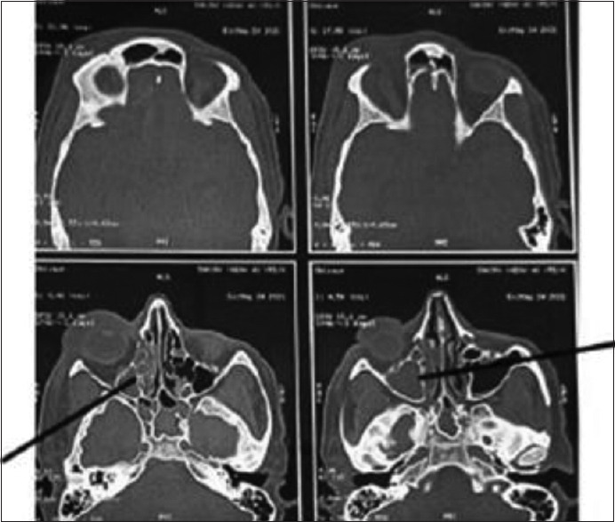
CT axial images of skull showing ill defined soft tissue density noted filling right maxillary, ethmoid and sphenoid sinus with rarefaction of anterior and medial wall of maxillary sinus with mild right orbital bulge and periorbital swelling
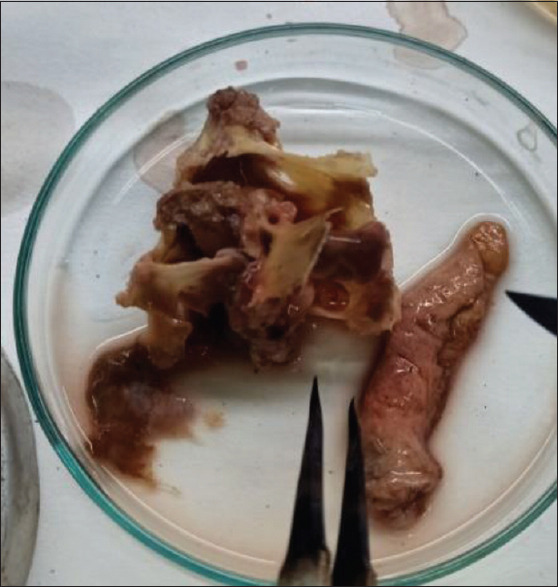
Excised tissue sample for microbiological investigation
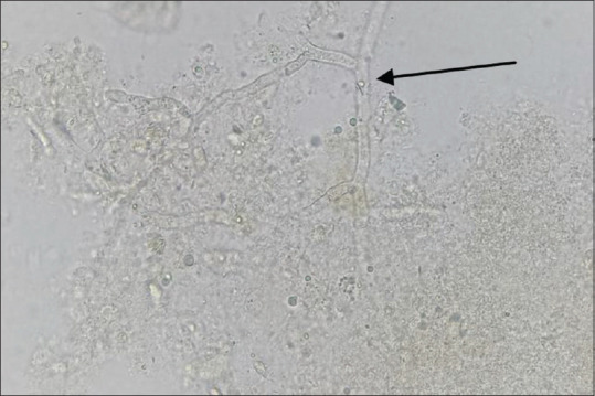
KOH wet mount of the excised tissue showing broad aseptate hyaline hyphae with right angle branching (Arrow), 400×
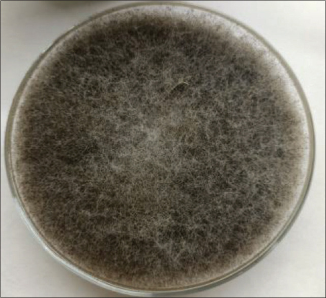
Cottony-fluffy growth of Rhizopus arrhizus on SDA agar
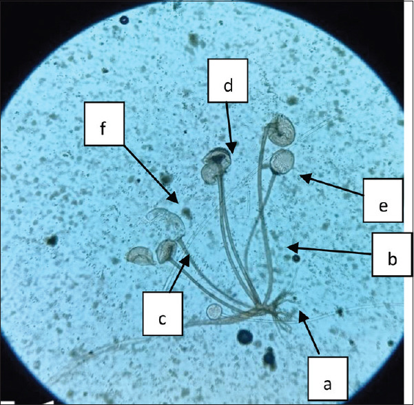
LPCB wet mount showing Rhizoid (a), sporangiophore (b), Apophysis (c), columella (d), collarets (e), sporangium (f), 400×
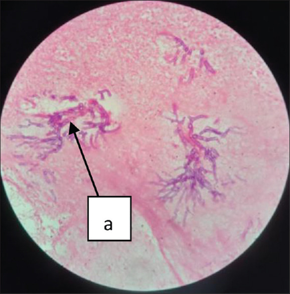
H and E staining of the excised tissue showing the necrosis (a) with plenty of broad aseptate hyphae (arrow), 400×
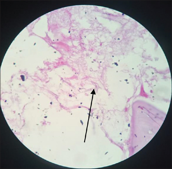
Pink coloured aseptate fungal hyphae (arrow), 400×
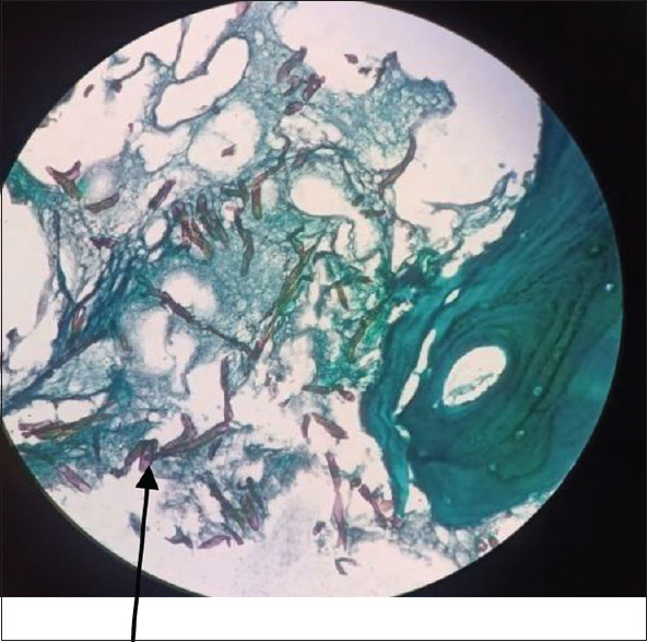
Dark brown-black coloured aspetate fungal hyphae (arrow), 400×
References
-
- Hibbett DS, Binder M, Bischoff JF, Blackwell M, Cannon PF, Eriksson OE, et al. A higher-level phylogenetic classification of the Fungi. Mycol Res. 2007;111:509–47. - PubMed
-
- Jeong W, Keighley C, Wolfe R, Lee WL, Slavin MA, Kong DC, et al. The epidemiology and clinical manifestations of mucormycosis:A systematic review and meta-analysis of case reports. Clin Microbiol Infect. 2019;25:26–34. - PubMed
-
- Richardson M. The ecology of the Zygomycetes and its impact on environmental exposure. Clin Microbiol Infect. 2009;15:2–9. - PubMed
Publication types
LinkOut - more resources
Full Text Sources
Miscellaneous