Left Ventricular Free Wall Rupture After Acute Myocardial Reinfarction Due to In-Stent Thrombosis in COVID-19 Patient
- PMID: 35800909
- PMCID: PMC9226759
- DOI: 10.5455/aim.2022.30.76-80
Left Ventricular Free Wall Rupture After Acute Myocardial Reinfarction Due to In-Stent Thrombosis in COVID-19 Patient
Abstract
Background: Acute left ventricular free wall rupture (LVFWR) is a life-threatening complication of myocardial infarction that requires urgent intervention. Surgical repair has continued to be the treatment of choice. Studies suggest a posterolateral or inferior infarction is more likely to result in free wall rupture than an anterior infarction. LVFWR generally results in death within minutes of the onset of recurrent chest pain, and on average was associated with a median survival time of 8 hours. Prompt diagnosis and management can lead to successful treatment for LVFWR.
Objective: The aim of this article was to present an emergency case with an LVFWR in a COVID-19 patient who suffers from AMI and was treated with PCI stents in the ramus intermedius and circumflex coronary artery.
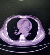
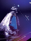
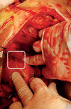
Case report: We present an emergency case with an LVFWR in a COVID-19 patient who suffers from AMI and was treated with PCI stents in the ramus intermedius and circumflex coronary artery. Although dual antiplatelet therapy introduction and good outcome of PCI were achieved, soon after instant thrombosis of both stents appear to result in transmural necrosis and LVFWR. Urgent catheterization was performed and diagnosed in-stent thrombosis where the ventriculography confirmed LVFWR of the posteroinferior wall. Urgent surgery was performed. Transmural necrosis was noticed alongside the incision line. The incision is sawn with 4 U-stitches (Prolen 2.0 with Teflon buttressed stitches). Another layer of fixation was made by Prolen 2.0 running stitches reinforced with Teflon felts from both sides. A large PTFE patch was fixed to epicardium over the suture line by Prolen 6.0 running stitch and BioGlue was injected in-between patch and LV (Figures 8 and 9). After aortic cross-clamp removal, the sinus rhythm was restored.
Conclusion: Despite the high mortality, the urgency and the complexity of surgical treatment the early diagnosis plays a key role in the management of postinfarction LVFWR patients presenting a case of preserved postoperative left ventricular function and accomplished good functional status, as presented in our case.
Keywords: COVID-19; acute myocardial infarction; left ventricular free wall rupture.
© 2022 Alen Karic, Ilirijana Haxhibeqiri-Karabdic, Edin Kabil, Sanja Grabovica, Slavenka Straus, Ervin Busevac, Alma Krajinovic, Bedrudin Banjanovic, Muhamed Djedovic, Nermir Granov.
Conflict of interest statement
There are no conflicts of interest.
Figures


References
-
- Glower DD. Cohn Lawrence H, Adams David H., editors. Surgical Treatment of Complications of Myocardial Infarction, Ventricular Septal Defect, Myocardial Rupture, and Left Ventricular Aneurysm. Cardiac Surgery in the Adult. (Fifth Edition) 2018:595–629. Copyright © 2018 by McGraw-Hill Education.
-
- Nishiyama K, Okino S, Andou J, et al. Coronary angioplasty reduces free wall rupture and improves mortality and morbidity of acute myocardial infarction. J Invasive Cardiol. 2004;16:554–548. - PubMed
-
- Graham JM, Feliciano DV, Mattox KL, Beall AC., Jr Innominate vascular injury. J Trauma. 1982;22:647–655. - PubMed
Publication types
LinkOut - more resources
Full Text Sources
Miscellaneous
