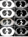
Solitary primary pulmonary synovial sarcoma: A case report
- PMID: 35801048
- PMCID: PMC9198894
- DOI: 10.12998/wjcc.v10.i15.5103
Solitary primary pulmonary synovial sarcoma: A case report
Abstract
Background: Synovial sarcoma (SS) is an uncommon and highly malignant soft tissue sarcoma in the clinic, with primary pulmonary SS (PPSS) being extremely rare. Here, we describe the clinical characteristics, diagnosis, and treatment of a solitary PPSS case confirmed via surgical resection and fluorescence in situ hybridization (FISH).
Case summary: A 33-year-old man was admitted because of intermittent coughing and hemoptysis for one month, with lung shadows observed for two years. Whole-body positron emission tomography-computed tomography (PET-CT) revealed a solitary mass in the upper lobe of the right lung, with uneven radioactivity uptake and a maximum standardized uptake value of 5.6. The greyish-yellow specimen obtained following thoracoscopic resection was covered with small multi-nodulated structures and consisted of soft tissue. Hematoxylin and eosin staining revealed spindle-shaped malignant tumor cells. Immunohistochemistry indicated these tumor cells were CD99 and BCL-2-positive. Furthermore, the FISH test revealed synovial sarcoma translocation genetic reassortment, which confirmed the diagnosis of SS.
Conclusion: PPSS is extremely rare and tends to be misdiagnosed as many primary pulmonary diseases. PET-CT, histologic analysis, and FISH tests can be used to differentiate PPSS from other diseases. Surgical resection is regularly recommended for the treatment of solitary PPSS and is helpful for improving the prognosis.
Keywords: Case report; Fluorescence in situ hybridization; Positron emission tomography-computed tomography; Primary pulmonary synovial sarcoma; Solitary pulmonary nodule; Spindle cells; Synovial sarcoma translocation.
©The Author(s) 2022. Published by Baishideng Publishing Group Inc. All rights reserved.
Conflict of interest statement
Conflict-of-interest statement: The authors declare that they have no conflict of interest.
Figures


References
-
- Mankin HJ, Hornicek FJ. Diagnosis, classification, and management of soft tissue sarcomas. Cancer Control. 2005;12:5–21. - PubMed
-
- Panigrahi MK, Pradhan G, Sahoo N, Mishra P, Patra S, Mohapatra PR. Primary pulmonary synovial sarcoma: A reappraisal. J Cancer Res Ther. 2018;14:481–489. - PubMed
-
- Zaborowski M, Vargas AC, Pulvers J, Clarkson A, de Guzman D, Sioson L, Maclean F, Chou A, Gill AJ. When used together SS18-SSX fusion-specific and SSX C-terminus immunohistochemistry are highly specific and sensitive for the diagnosis of synovial sarcoma and can replace FISH or molecular testing in most cases. Histopathology. 2020;77:588–600. - PubMed
-
- Suurmeijer AJH, de Bruijn D, Geurts van Kessel A, Miettinen MM. Synovial sarcoma. In: Fletcher CDM, Bridge JA, Hogendoor P, Mertens F, editing. WHO classification of soft tissue and bone tumors. Fourth edition. Lyon: IARC. 2013: 213-215.
-
- Etienne-Mastroianni B, Falchero L, Chalabreysse L, Loire R, Ranchère D, Souquet PJ, Cordier JF. Primary sarcomas of the lung: a clinicopathologic study of 12 cases. Lung Cancer. 2002;38:283–289. - PubMed
Publication types
LinkOut - more resources
Full Text Sources

