

The Role of Mesenchymal Stem Cells in Regulating Astrocytes-Related Synapse Dysfunction in Early Alzheimer's Disease
- PMID: 35801178
- PMCID: PMC9253587
- DOI: 10.3389/fnins.2022.927256
The Role of Mesenchymal Stem Cells in Regulating Astrocytes-Related Synapse Dysfunction in Early Alzheimer's Disease
Abstract
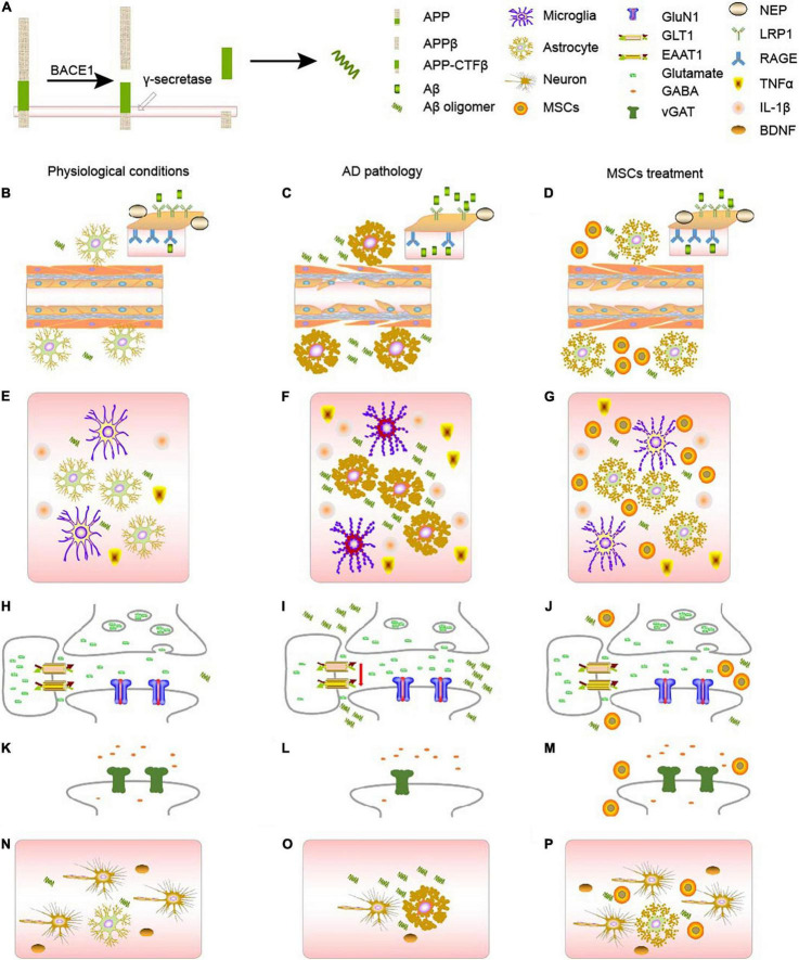
Alzheimer's disease (AD), a neurodegenerative disease, is characterized by the presence of extracellular amyloid-β (Aβ) aggregates and intracellular neurofibrillary tangles formed by hyperphosphorylated tau as pathological features and the cognitive decline as main clinical features. An important cellular correlation of cognitive decline in AD is synapse loss. Soluble Aβ oligomer has been proposed to be a crucial early event leading to synapse dysfunction in AD. Astrocytes are crucial for synaptic formation and function, and defects in astrocytic activation and function have been suggested in the pathogenesis of AD. Astrocytes may contribute to synapse dysfunction at an early stage of AD by participating in Aβ metabolism, brain inflammatory response, and synaptic regulation. While mesenchymal stem cells can inhibit astrogliosis, and promote non-reactive astrocytes. They can also induce direct regeneration of neurons and synapses. This review describes the role of mesenchymal stem cells and underlying mechanisms in regulating astrocytes-related Aβ metabolism, neuroinflammation, and synapse dysfunction in early AD, exploring the open questions in this field.
Keywords: Alzheimer’s disease; amyloid β; astrocytes; mesenchymal stem cells; neuroinflammation; synaptic functions.
Copyright © 2022 Liu.
Conflict of interest statement
The author declares that the research was conducted in the absence of any commercial or financial relationships that could be construed as a potential conflict of interest.
Figures
Similar articles
-
Synaptic Mitochondria: An Early Target of Amyloid-β and Tau in Alzheimer's Disease.J Alzheimers Dis. 2021;84(4):1391-1414. doi: 10.3233/JAD-215139. J Alzheimers Dis. 2021. PMID: 34719499 Review.
-
Mechanism of Oxidative Stress and Synapse Dysfunction in the Pathogenesis of Alzheimer's Disease: Understanding the Therapeutics Strategies.Mol Neurobiol. 2016 Jan;53(1):648-661. doi: 10.1007/s12035-014-9053-6. Epub 2014 Dec 17. Mol Neurobiol. 2016. PMID: 25511446 Free PMC article. Review.
-
The Role of Amyloid-Beta and Tau in the Early Pathogenesis of Alzheimer's Disease.Med Sci Monit. 2021 Sep 2;27:e933084. doi: 10.12659/MSM.933084. Med Sci Monit. 2021. PMID: 34471085 Free PMC article. Review.
-
Defective mitophagy and synaptic degeneration in Alzheimer's disease: Focus on aging, mitochondria and synapse.Free Radic Biol Med. 2021 Aug 20;172:652-667. doi: 10.1016/j.freeradbiomed.2021.07.013. Epub 2021 Jul 8. Free Radic Biol Med. 2021. PMID: 34246776 Review.
-
Urokinase-Type Plasminogen Activator Protects Cerebral Cortical Neurons from Soluble Aβ-Induced Synaptic Damage.J Neurosci. 2020 May 20;40(21):4251-4263. doi: 10.1523/JNEUROSCI.2804-19.2020. Epub 2020 Apr 24. J Neurosci. 2020. PMID: 32332118 Free PMC article.
Cited by
-
Therapeutic potential of mesenchymal stem cells for cerebral small vessel disease.Regen Ther. 2024 Feb 22;25:377-386. doi: 10.1016/j.reth.2023.11.002. eCollection 2024 Mar. Regen Ther. 2024. PMID: 38414558 Free PMC article. Review.
-
Mesenchymal Stem Cells Applications in Alzheimer's Disease.Glob Med Genet. 2023 Dec 11;10(4):382-387. doi: 10.1055/s-0043-1777087. eCollection 2023 Dec. Glob Med Genet. 2023. PMID: 38089680 Free PMC article. Review.
-
Emerging Role of Mesenchymal Stromal Cell and Exosome Therapies in Treating Cognitive Impairment.Pharmaceutics. 2025 Feb 20;17(3):284. doi: 10.3390/pharmaceutics17030284. Pharmaceutics. 2025. PMID: 40142948 Free PMC article. Review.
-
Aβ-mediated synaptic glutamate dynamics and calcium dynamics in astrocytes associated with Alzheimer's disease.Cogn Neurodyn. 2024 Dec;18(6):3401-3426. doi: 10.1007/s11571-024-10064-6. Epub 2024 Feb 17. Cogn Neurodyn. 2024. PMID: 39712135
-
Use of Brain-Derived Stem/Progenitor Cells and Derived Extracellular Vesicles to Repair Damaged Neural Tissues: Lessons Learned from Connective Tissue Repair Regarding Variables Limiting Progress and Approaches to Overcome Limitations.Int J Mol Sci. 2023 Feb 8;24(4):3370. doi: 10.3390/ijms24043370. Int J Mol Sci. 2023. PMID: 36834779 Free PMC article. Review.
References
-
- Blasko I., Veerhuis R., Stampfer-Kountchev M., Saurwein-Teissl M., Eikelenboom P., Grubeck-Loebenstein B. (2000). Costimulatory effects of interferon-gamma and interleukin-1beta or tumor necrosis factor alpha on the synthesis of Abeta1-40 and Abeta1-42 by human astrocytes. Neurobiol. Dis. 7 682–689. 10.1006/nbdi.2000.0321 - DOI - PubMed
Publication types
LinkOut - more resources
Full Text Sources