

Metabolic arsenal of giant viruses: Host hijack or self-use?
- PMID: 35801640
- PMCID: PMC9270025
- DOI: 10.7554/eLife.78674
Metabolic arsenal of giant viruses: Host hijack or self-use?
Abstract
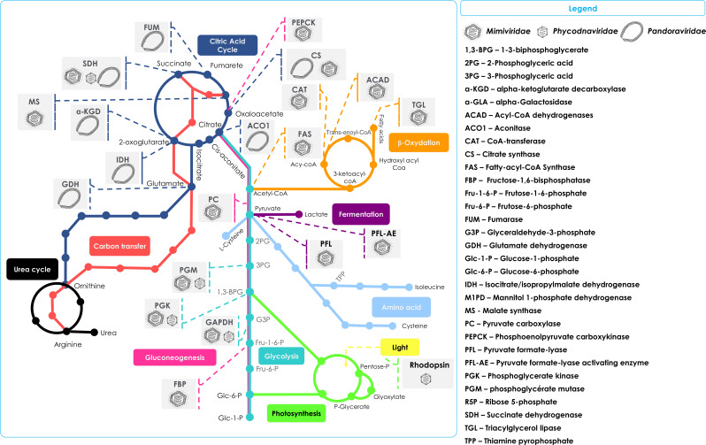
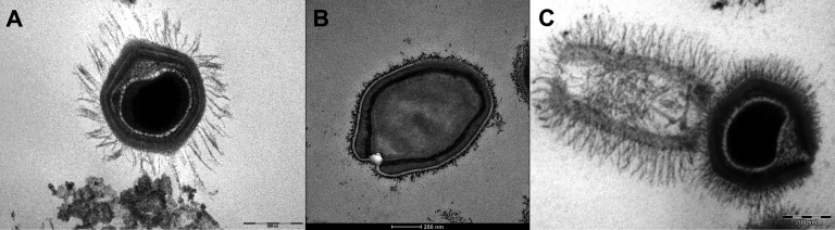
Viruses generally are defined as lacking the fundamental properties of living organisms in that they do not harbor an energy metabolism system or protein synthesis machinery. However, the discovery of giant viruses of amoeba has fundamentally challenged this view because of their exceptional genome properties, particle sizes and encoding of the enzyme machinery for some steps of protein synthesis. Although giant viruses are not able to replicate autonomously and still require a host for their multiplication, numerous metabolic genes involved in energy production have been recently detected in giant virus genomes from many environments. These findings have further blurred the boundaries that separate viruses and living organisms. Herein, we summarize information concerning genes and proteins involved in cellular metabolic pathways and their orthologues that have, surprisingly, been discovered in giant viruses. The remarkable diversity of metabolic genes described in giant viruses include genes encoding enzymes involved in glycolysis, gluconeogenesis, tricarboxylic acid cycle, photosynthesis, and β-oxidation. These viral genes are thought to have been acquired from diverse biological sources through lateral gene transfer early in the evolution of Nucleo-Cytoplasmic Large DNA Viruses, or in some cases more recently. It was assumed that viruses are capable of hijacking host metabolic networks. But the giant virus auxiliary metabolic genes also may represent another form of host metabolism manipulation, by expanding the catalytic capabilities of the host cells especially in harsh environments, providing the infected host cells with a selective evolutionary advantage compared to non-infected cells and hence favoring the viral replication. However, the mechanism of these genes' functionality remains unclear to date.
Keywords: energy production; giant viruses; infectious disease; microbiology; primary metabolism.
© 2022, Belhaouari et al.
Conflict of interest statement
DB, GP, DL, SK, JG, JS, PC, BL, SA No competing interests declared
Figures
References
-
- Abrahão J, Silva L, Silva LS, Khalil JYB, Rodrigues R, Arantes T, Assis F, Boratto P, Andrade M, Kroon EG, Ribeiro B, Bergier I, Seligmann H, Ghigo E, Colson P, Levasseur A, Kroemer G, Raoult D, La Scola B. Tailed giant Tupanvirus possesses the most complete translational apparatus of the known virosphere. Nature Communications. 2018;9:1–12. doi: 10.1038/s41467-018-03168-1. - DOI - PMC - PubMed
-
- Aherfi S, Andreani J, Baptiste E, Oumessoum A, Dornas FP, Andrade A, Chabriere E, Abrahao J, Levasseur A, Raoult D, La Scola B, Colson P. A Large Open Pangenome and A Small Core Genome for Giant Pandoraviruses. Frontiers in Microbiology. 2018;9:1486. doi: 10.3389/fmicb.2018.01486. - DOI - PMC - PubMed
-
- Aherfi S, Brahim Belhaouari D, Pinault L, Baudoin J-P, Decloquement P, Abrahao J, Colson P, Levasseur A, Lamb DC, Chabriere E, Raoult D, La Scola B. Incomplete tricarboxylic acid cycle and proton gradient in Pandoravirus massiliensis: is it still a virus? The ISME Journal. 2022;16:695–704. doi: 10.1038/s41396-021-01117-3. - DOI - PMC - PubMed
Publication types
MeSH terms
Grants and funding
LinkOut - more resources
Full Text Sources
