

Systemic calcinosis in a Quarter Horse gelding homozygous for a myosin heavy chain 1 mutation
- PMID: 35801821
- PMCID: PMC9308413
- DOI: 10.1111/jvim.16481
Systemic calcinosis in a Quarter Horse gelding homozygous for a myosin heavy chain 1 mutation
Abstract
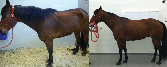
Case description: A 9-year-old Quarter Horse gelding was presented for lethargy, decreased appetite, polyuria and polydipsia (PU/PD), and severe muscle wasting suggestive of immune-mediated myositis.
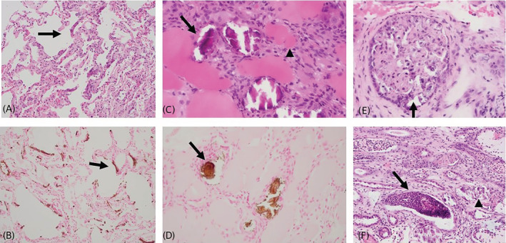
Clinical findings: The horse displayed lethargy, fever, tachyarrhythmia, inappetence, PU/PD, and severe epaxial and gluteal muscle wasting. Clinicopathologic findings were consistent with previously reported cases of systemic calcinosis in horses, including increased muscle enzyme activity, hyperphosphatemia, increased calcium-phosphorus product, hypoproteinemia, and an inflammatory leukogram. A diagnosis of systemic calcinosis was established by histopathologic evaluation of biopsy specimens from skeletal muscle, lung, and kidney.
Treatment and outcome: Symptomatic treatment was complemented by IV treatment with sodium thiosulfate to reverse calcium-phosphate precipitation in soft tissue and PO aluminum hydroxide to decrease intestinal phosphorus absorption and serum phosphorus concentration.
Clinical relevance: This is the first report in the veterinary literature of an antemortem diagnosis of systemic calcinosis in the horse that was successfully treated and had favorable long-term outcome.
Keywords: calcium-phosphorus product; immune mediated myositis; polydipsia; polyuria; sodium thiosulfate.
© 2022 The Authors. Journal of Veterinary Internal Medicine published by Wiley Periodicals LLC. on behalf of the American College of Veterinary Internal Medicine.
Conflict of interest statement
Authors declare no conflict of interest.
Figures


Similar articles
-
Myosin heavy-chain myopathy in 2 American quarter horses.Vet Pathol. 2024 May;61(3):462-467. doi: 10.1177/03009858231204253. Epub 2023 Oct 11. Vet Pathol. 2024. PMID: 37818977
-
Systemic calcinosis in horses: Pathological and genetic aspects.Equine Vet J. 2025 Jul;57(4):1017-1027. doi: 10.1111/evj.14464. Epub 2025 Jan 6. Equine Vet J. 2025. PMID: 39763093
-
Suspected systemic calcinosis and calciphylaxis in 5 horses.Can Vet J. 2010 Sep;51(9):993-9. Can Vet J. 2010. PMID: 21119866 Free PMC article.
-
Immune-Mediated Muscle Diseases of the Horse.Vet Pathol. 2018 Jan;55(1):68-75. doi: 10.1177/0300985816688755. Epub 2017 Jan 27. Vet Pathol. 2018. PMID: 28129093 Review.
-
Myosin Heavy Chain Myopathy and Immune-Mediated Muscle Disorders.Vet Clin North Am Equine Pract. 2025 Apr;41(1):61-75. doi: 10.1016/j.cveq.2024.10.005. Epub 2025 Jan 28. Vet Clin North Am Equine Pract. 2025. PMID: 39880733 Review.
References
-
- Fales‐Williams A, Sponseller B, Flaherty H. Idiopathic arterial medial calcification of the thoracic arteries in adult horses. J Vet Diagn Invest. 2008;20:692‐697. - PubMed
-
- Durward‐Akhurst S, Valberg SJ. Inflammatory and immune‐mediated muscle disorders. In: Felippe JB, ed. Equine Clinical Immunology. 1st ed. New York, NY: Wiley‐Blackwell; 2016:91‐99.
-
- Durward‐Akhurst SA, Valberg SJ. Immune‐mediated muscle diseases of the horse. Vet Pathol. 2018;55:68‐75. - PubMed
Publication types
MeSH terms
Substances
LinkOut - more resources
Full Text Sources
Medical
