. 2022 Jul;70(7):515-530.
doi: 10.1369/00221554221112702. Epub 2022 Jul 8.

Increased Colocalization and Interaction Between Decidual Protein Kinase A and Insulin-like Growth Factor-Binding Protein-1 in Intrauterine Growth Restriction

Affiliations

Increased Colocalization and Interaction Between Decidual Protein Kinase A and Insulin-like Growth Factor-Binding Protein-1 in Intrauterine Growth Restriction

Madhulika B Gupta et al. J Histochem Cytochem. 2022 Jul.

Abstract

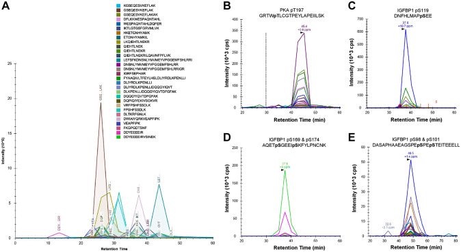
Increased phosphorylation of decidual insulin-like growth factor-binding protein-1 (IGFBP-1) can contribute to intrauterine growth restriction (IUGR) by decreasing the bioavailability of insulin-like growth factor-1 (IGF-1). However, the molecular mechanisms regulating IGFBP-1 phosphorylation at the maternal-fetal interface are poorly understood. Protein kinase A (PKA) is required for normal decidualization. Consensus sequences for PKA are present in IGFBP-1. We hypothesized that the expression/interaction of PKA with decidual IGFBP-1 is increased in IUGR. Parallel reaction monitoring-mass spectrometry (PRM-MS) identified multiple PKA peptides (n=>30) co-immunoprecipitating with IGFBP-1 in decidualized primary human endometrial stromal cells (HESC). PRM-MS also detected active PKApThr197 and greater site-specific IGFBP-1 phosphorylation(pSer119), (pSer98+pSer101) (pSer169+pSer174) in response to hypoxia. Hypoxia promoted colocalization [dual immunofluorescence (IF)] of PKA with IGFBP-1 in decidualized HESC. Colocalization (IF) and interaction (proximity ligation assay) of PKA and IGFBP-1 were increased in decidua collected from placenta of human IUGR pregnancies (n=8) compared with decidua from pregnancies with normal fetal growth. Similar changes were detected in decidual PKA/IGFBP-1 using placenta from baboons subjected to maternal nutrient reduction (MNR) vs controls (n=3 each). In baboons, these effects were evident in MNR at gestational day 120 prior to IUGR onset. Increased PKA-mediated phosphorylation of decidual IGFBP-1 may contribute to decreased IGF-1 bioavailability in the maternal-fetal interface in IUGR.

Keywords: fluorescent antibody technique; humans; hypoxia; mass spectrometry; maternal; nutrient reduction; papio; placenta; stromal cells.


Conflict of interest statement

Competing Interests: The author(s) declared no potential conflicts of interest with respect to the research, authorship, and/or publication of this article.

Figures

Figure 1.
Figure 1.
PRM-MS led to the identification of PKA-specific peptides from IGFBP-1 IP. Detection of PKA peptides by PRM-MS from IGFBP-1 immunoprecipitation (A). Chromatogram shows total transitions for 30 PKA-derived peptides; each peak monitors at least four daughter ions. Representative spectra of each daughter ion used to detect the presence of active PKC-pThr197 from IP samples (B). Codetection of IGFBP-1 phosphorylated peptides (C–E). Chromatographs show transitions for four IGFBP-1 peptides, IGFBP1-pSer119 (C), doubly phosphorylated IGFBP-1-pSer196 + pSer174 (D), and doubly phosphorylated IGFBP-1-pSer98 + pSer174 (E). All samples were doubly digested by trypsin and Asp-N protease and C18 cleaned prior to MS. Abbreviations: PRM-MS, parallel reaction monitoring-mass spectrometry; PKA, protein kinase A; IGFBP-1, insulin-like growth factor binding protein-1; IP, immunoprecipitation.
Figure 2.
Figure 2.
PRM-MS of relative PKA and IGFBP-1 phosphorylation in HESC samples in normoxic and hypoxic conditions. Active PKA autophosphorylation at Thr197 (A); IGFBP-1 phosphorylation at Ser119, Ser98+Ser101, and Ser169 + Ser174 (B). Relative values are determined from total peak intensities of transition ions from PRM-MS and normalized for protein abundance using internal peptide controls. Abbreviations: PRM-MS, parallel reaction monitoring-mass spectrometry; PKA, protein kinase A; IGFBP-1, insulin-like growth factor–binding protein-1; HESC, human endometrial stromal cells.
Figure 3.
Figure 3.
IGFBP-1 and PKA colocalization increases in decidualized T-HESC in response to hypoxia. Representative dual immunofluorescence staining of IGFBP-1 (red) (A, E) and protein kinase PKA (green) (B, F) in decidualized HESC cultured in normoxia or hypoxia. Merged confocal images depict colocalization between IGFBP-1 and PKA (red and green combined yield yellow) for normoxia and hypoxia (C, G). Nuclei are stained with DAPI (blue) (D, H). Scale bar = 25 µm. Abbreviations: IGFBP-1, insulin-like growth factor–binding protein-1; PKA, protein kinase A; DAPI, 4′,6-diamidino-2-phenylindole; HESC, transformed human endometrial stromal cells.
Figure 4.
Figure 4.
Increased colocalization of decidual IGFBP-1 and PKA in term human IUGR placenta. Representative immunofluorescence images of IGFBP-1 (Alexa 568, red) and protein kinase A (Alexa 660, green) are shown in AGA and from the GA-matched IUGR placenta. Images were captured across the entire decidual area for each placenta, which subsequently provided an adequately large number of images (n=12) to minimize bias. Both IGFBP-1 (red) and PKA (green) signals along with single channel images (A–D) are shown with increased signals in the decidual region of the placenta. Merged multichannel images indicate colocalized signal (C–F). Nuclei are stained with DAPI (blue) (A–B); negative control (G). Original scale bars, 20 µM. Abbreviations: IGFBP-1, insulin-like growth factor–binding protein-1; PKA, protein kinase A; IUGR, intrauterine growth restriction; DAPI, 4′,6-diamidino-2-phenylindole; GA, gestational age; AGA, appropriate for gestational age.
Figure 5.
Figure 5.
Increased colocalization of decidual IGFBP-1 and PKA in term human IUGR placenta as demonstrated by PLA. Representative PLA signals in images of IGFBP-1 and PKA in the decidual region of human placenta (indicated by the red dots) demonstrate the potential interaction in IUGR compared with AGA. Cropped and zoomed-in images (C–D) correspond to the white-dashed boxes in (A–B). Nuclei are stained with DAPI (blue). Negative controls (E–F). Bar graph indicates an increase in PLA signals in IUGR compared with AGA (p=0.006). Original scale bars, 50 µM. Abbreviations: IGFBP-1, insulin-like growth factor–binding protein-1; PKA, protein kinase A; IUGR, intrauterine growth restriction; PLA, proximity ligation assay; DAPI, 4′,6-diamidino-2-phenylindole; AGA, appropriate for gestational age.
Figure 6.
Figure 6.
Increased decidual IGFBP-1 and vimentin colocalization in MNR baboons at gestational day 120. Representative immunofluorescence staining of IGFBP-1 (green) and vimentin (red) is shown in control and MNR baboon placenta at gestational day 120. Decidual region shows increased colocalization of IGFBP-1 and vimentin (indicated in yellow) in MNR (F) compared with control (C) in merged channels. Negative controls (G and H). Nuclei are stained with DAPI (blue). Abbreviations: IGFBP-1, insulin-like growth factor binding protein-1; MNR, maternal nutrient reduction; DAPI, 4′,6-diamidino-2-phenylindole; IUGR, intrauterine growth restriction; PLA, proximity ligation assay; AGA, appropriate for gestational age.
Figure 7.
Figure 7.
Increased decidual IGFBP-1 and vimentin colocalization in MNR baboons at gestational day 165. Representative immunofluorescence staining of IGFBP-1 (green) and vimentin (red) in control and MNR baboon placenta at gestational day 165. Decidual region shows increased colocalization of IGFBP-1 and vimentin (indicated in yellow) in MNR placenta (F) compared with control (C) in merged channel images. Negative controls (G and H). Original scale bars, 50 µM. Abbreviations: IGFBP-1, insulin-like growth factor binding protein-1; MNR, maternal nutrient reduction; DAPI, 4′,6-diamidino-2-phenylindole.
Figure 8.
Figure 8.
Increased decidual IGFBP-1 and PKA colocalization in MNR baboons at gestational day 120. Representative dual immunofluorescence staining of PKA (green) and IGFBP-1 (red) in control and MNR baboon placenta at gestational day 120. Decidual region shows increased colocalization of PKA and IGFBP-1 (indicated in yellow) in MNR (F) compared with control (C) in merged channel images. Negative controls (G and H). Nuclei are stained with DAPI (blue). Abbreviations: IGFBP-1, insulin-like growth factor–binding protein-1; PKA, protein kinase A; MNR, maternal nutrient reduction; DAPI, 4′,6-diamidino-2-phenylindole.
Figure 9.
Figure 9.
Increased decidual IGFBP-1 and PKA colocalization in MNR baboons at gestational day 120 as shown by deconvolution of images. Representative immunofluorescence staining of PKA (green) and IGFBP-1 (red) in the decidual region of control and MNR baboon placenta is shown by two-dimensional deconvolution of images. Single-channel IGFBP-1 (A and D) and PKA (B and E) are shown as multichannel (C) and (F) deconvolved images indicating colocalization of IGFBP-1 and PKA in the decidua of control and MNR baboons, with marked increase in areas of colocalization in the MNR decidua. Abbreviations: IGFBP-1, insulin-like growth factor–binding protein-1; PKA, protein kinase A; MNR, maternal nutrient reduction; DAPI, 4′,6-diamidino-2-phenylindole; AGA, appropriate for gestational age.
Figure 10.
Figure 10.
Increased decidual IGFBP-1 and PKA colocalization in MNR baboons at gestational day 165. Representative immunofluorescence staining of PKA (green) and IGFBP-1 (red) in the decidual region of control and MNR baboon placenta at gestational day 165. Decidual region shows increased colocalization of PKA and IGFBP-1 (indicated in yellow) in MNR (G) compared with control (C) in merged channel images. Negative controls (H and I). Nuclei are stained with DAPI (blue). Abbreviations: IGFBP-1, insulin-like growth factor binding protein-1; PKA, Protein kinase A; MNR, maternal nutrient reduction; DAPI, 4′,6-diamidino-2-phenylindole; AGA, appropriate for gestational age.
Figure 11.
Figure 11.
Increased decidual IGFBP-1 and PKA colocalization in MNR baboons at gestational days 120 and 165 as demonstrated by PLA. PLA signals in representative images demonstrate the potential interaction of IGFBP-1 and PKA in the decidual region of baboon placenta tissue (indicated by the red dots) and a greater interaction in MNR compared with control. Placentas from GD120 (control and MNR n=5 each) and GD165 baboons (control and MNR n=3 each) were used. Cropped and zoomed images (C–D) correspond to the white-dashed boxes in (A–B). Cropped and zoomed images (I–J) correspond to the white-dashed boxes in (G–H). Nuclei are stained with DAPI (blue). Respective negative controls are shown. Abbreviations: IGFBP-1, insulin-like growth factor–binding protein-1; PKA, protein kinase A; MNR, maternal nutrient reduction; PLA, proximity ligation assay; DAPI, 4′,6-diamidino-2-phenylindole; IUGR, intrauterine growth restriction.

Similar articles

Cited by

References

    1. Gibson JM, Aplin JD, White A, Westwood M. Regulation of IGF bioavailability in pregnancy. Mol Hum Reprod. 2001;7(1):79–87. - PubMed
    1. Clemmons DR. IGF binding proteins and their functions. Mol Reprod Dev. 1993;35(4):368–74. - PubMed
    1. Martina NA, Kim E, Chitkara U, Wathen NC, Chard T, Giudice LC. Gestational age-dependent expression of insulin-like growth factor-binding protein-1 (IGFBP-1) phosphoisoforms in human extraembryonic cavities, maternal serum, and decidua suggests decidua as the primary source of IGFBP-1 in these fluids during early pregnancy. J Clin Endocrinol Metab. 1997;82(6):1894–8. - PubMed
    1. Seferovic MD, Ali R, Kamei H, Liu S, Khosravi JM, Nazarian S, Han VK, Duan C, Gupta MB. Hypoxia and leucine deprivation induce human insulin-like growth factor binding protein-1 hyperphosphorylation and increase its biological activity. Endocrinology. 2009; 150(1):220–31. - PMC - PubMed
    1. Westwood M, Gibson JM, Davies AJ, Young RJ, White A. The phosphorylation pattern of insulin-like growth factor-binding protein-1 in normal plasma is different from that in amniotic fluid and changes during pregnancy. J Clin Endocrinol Metab. 1994;79(6):1735–41. - PubMed

Publication types

Substances