. 2022 Sep 2;82(17):3158-3171.
doi: 10.1158/0008-5472.CAN-22-0170.

Retinoic Acid Receptor Activation Reduces Metastatic Prostate Cancer Bone Lesions by Blocking the Endothelial-to-Osteoblast Transition

Affiliations

Retinoic Acid Receptor Activation Reduces Metastatic Prostate Cancer Bone Lesions by Blocking the Endothelial-to-Osteoblast Transition

Guoyu Yu et al. Cancer Res. .

Abstract

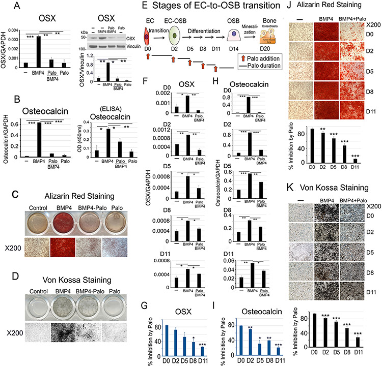
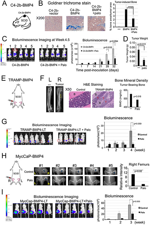
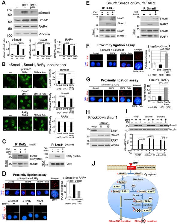
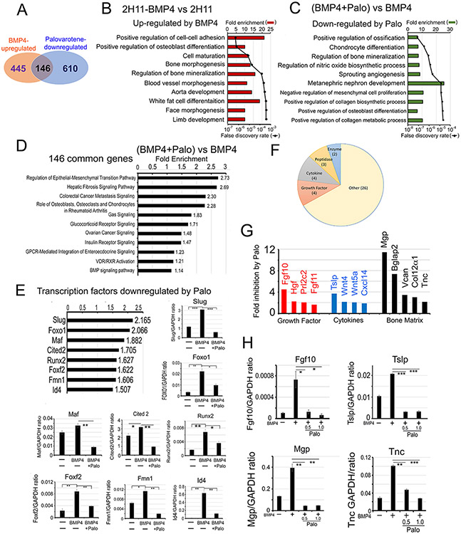
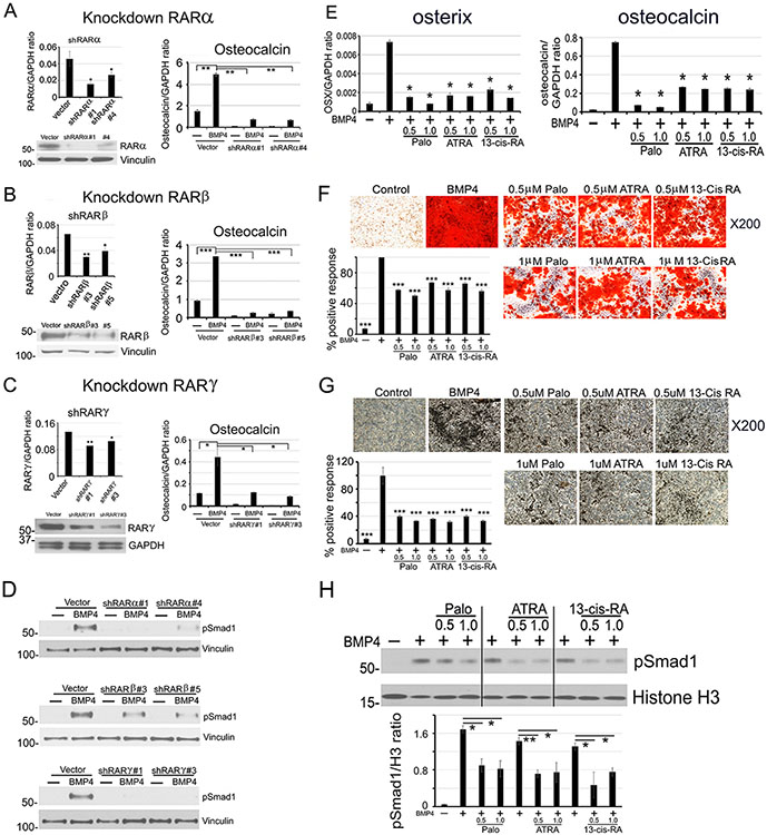
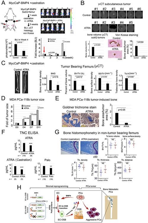
Metastatic prostate cancer in the bone induces bone-forming lesions that contribute to progression and therapy resistance. Prostate cancer-induced bone formation originates from endothelial cells (EC) that have undergone endothelial-to-osteoblast (EC-to-OSB) transition in response to tumor-secreted BMP4. Current strategies targeting prostate cancer-induced bone formation are lacking. Here, we show that activation of retinoic acid receptor (RAR) inhibits EC-to-OSB transition and reduces prostate cancer-induced bone formation. Treatment with palovarotene, an RARγ agonist being tested for heterotopic ossification in fibrodysplasia ossificans progressiva, inhibited EC-to-OSB transition and osteoblast mineralization in vitro and decreased tumor-induced bone formation and tumor growth in several osteogenic prostate cancer models, and similar effects were observed with the pan-RAR agonist all-trans-retinoic acid (ATRA). Knockdown of RARα, β, or γ isoforms in ECs blocked BMP4-induced EC-to-OSB transition and osteoblast mineralization, indicating a role for all three isoforms in prostate cancer-induced bone formation. Furthermore, treatment with palovarotene or ATRA reduced plasma Tenascin C, a factor secreted from EC-OSB cells, which may be used to monitor treatment response. Mechanistically, BMP4-activated pSmad1 formed a complex with RAR in the nucleus of ECs to activate EC-to-OSB transition. RAR activation by palovarotene or ATRA caused pSmad1 degradation by recruiting the E3-ubiquitin ligase Smad ubiquitination regulatory factor1 (Smurf1) to the nuclear pSmad1/RARγ complex, thus blocking EC-to-OSB transition. Collectively, these findings suggest that palovarotene can be repurposed to target prostate cancer-induced bone formation to improve clinical outcomes for patients with bone metastasis.

Significance: This study provides mechanistic insights into how RAR agonists suppress prostate cancer-induced bone formation and offers a rationale for developing RAR agonists for prostate cancer bone metastasis therapy. See related commentary by Bhowmick and Bhowmick, p. 2975.


Conflict of interest statement

Conflict of interest

C. J. Logothetis reports receiving commercial research grants from Janssen, ORIC Pharmaceuticals, Novartis, Aragon Pharmaceuticals; and honoraria from Merck, Sharp & Dohme, Bayer, Amgen. No potential conflicts of interest are disclosed by the other authors.

Figures

Figure 1.
Figure 1.. Palovarotene inhibits BMP4-induced EC-to-OSB transition in 2H11 cells.
(A-D) 2H11 ECs were treated with BMP4 (100 ng/mL) to induce EC-to-OSB transition. Palovarotene (Palo, 1 μM) effects were measured on (A) OSX mRNA (qRT-PCR) and protein (blot), (B) osteocalcin mRNA and protein (ELISA of conditioned medium), and mineralization by (C) Alizarin Red and (D) von Kossa staining. (E) Stages of EC-to-OSB transition. Red arrows, Palo addition at different times followed by a 3-day incubation. (F) OSX mRNA expression when Palo was added at D0, D2, D5, D8 or D11. (G) Percent inhibition of OSX expression by Palo in cells in (F) when compared to BMP4-treated only, using the formula 100-[(BMP4+Palo)/BMP4]x100. (H) Osteocalcin mRNA expression in cells treated as in (F). (I) Percent inhibition of osteocalcin expression by Palo as in (G). (J) Palo on mineralization when added at indicated days, as determined by Alizarin Red or (K) von Kossa staining. Staining quantified using ImageJ. *p<0.05, **p<0.01, ***p<0.001 by Students t-test in this and subsequent figures.
Figure 2.
Figure 2.. Palovarotene reduces PCa-induced bone formation and tumor growth in SCID mice.
(A) C4-2b-BMP4 subcutaneous implantation. (B) Goldner Trichrome staining of C4-2b-BMP4 tumors quantified using ImageJ. (C) Tumor size measurement by BLI in mice with C4-2b-BMP4 tumors. (D) Tumor weight at the termination of the study. Control (n=5), Palo-treated (n=5). (E) TRAMP-BMP4 intrabone injection. (F) MicroCT of femurs with or without TRAMP-BMP4 tumors (left). Histology of TRAMP-BMP4-induced bone compared with the non-tumor bearing bone (middle). B(bone), T(tumor). Palo on bone mineral density of tumor-bearing femurs (right). (G) TRAMP-BMP4 tumor size by BLI. Control (n=7); Palo-treated (n=5). (H) MycCaP-BMP4 intrabone injection (left). X-Ray of osteoblastic bone response induced by MycCaP-BMP4 in femurs with or without Palo (middle) and relative intensity quantified by ImageJ (right). Blue arrows, tumor bearing bone in the control group. (I) MycCaP-BMP4 tumor size by BLI. Control (n=5); Palo-treated (n=5).
Figure 3.
Figure 3.. Palovarotene decreases BMP4-stimulated pSmad1.
(A) pSmad1, Smad1 and RARγ protein levels in 2H11 cells treated as indicated for 6 h. Vinculin, loading control. Blots quantified by ImageJ. (B) Immunofluorescence of pSmad1, Smad1, and RARγ in nucleus of cells in (A). Relative intensity quantified by ImageJ. n, number of nuclei examined. All bars, 20 μm. (C) Co-immunoprecipitation of RARγ and pSmad1 from nuclear extracts. (D) Proximity ligation assay for the interaction of RARγ and pSmad1. Red spots, PLA signals in the nucleus using anti-Smad1 and anti-RARγ antibodies. Single or no antibodies were used as controls. (E) Co-immunoprecipitations of Smurf1 with RARγ or pSmad1 using nuclear extracts. Note in (C, E), efficiency of immunoprecipitations could not be assessed due to RARγ and Smad1 being similar in size as IgG bands. (F-G) PLA for Smurf1 with pSmad1 (F) or RARγ (G). (H) Smurf1 knockdown in 2H11-shSmurf1#1 or #3 clones. (I) pSmad1 levels in 2H11-shSmurf1 clones. (J) Graphical summary. BMP4 stimulates an increase in pSmad1 that translocates into the nucleus, where pSmad1 and RARγ form a complex. Palo results in the recruitment of E3-ubiquitin ligase Smurf1 to the pSmad1/RARγ complex, leading to pSmad1 degradation and inhibition of EC-to-OSB transition.
Figure 4.
Figure 4.. Palovarotene alters the BMP4-induced transcriptome.
(A) Venn diagram of number of genes whose expression levels are upregulated by BMP4 and downregulated by Palo in 2H11 cells. Pathway analyses of genes upregulated by BMP4 (B), downregulated by Palo after 48 h (C), and up-regulated by BMP4 but down-regulated by Palo (Palo-regulated genes) (D). (E) Transcription factors upregulated by BMP4 and downregulated by Palo. qRT-PCR for indicated mRNAs. (F) Category of BMP4-inducible secreted proteins that are inhibited by Palo. (G) BMP4-stimulated secreted proteins (termed “osteocrines”) (12) that are decreased by Palo. (H) qRT-PCR for select mRNAs in (G) treated as indicated.
Figure 5.
Figure 5.. All three RAR isoforms are involved in EC-to-OSB transition.
(A-C) (Left) RARα, RARβ, or RARγ knockdown in 2H11 cells by shRNA (qRT-PCR and western blot). (Right) RAR knockdowns on BMP4-stimulated osteocalcin expression (qRT-PCR). (D) RAR knockdowns on BMP4-stimulated nuclear pSmad1 at 48 h. (E-G) ATRA or 13-cis-RA on BMP4-stimulated osterix and osteocalcin expression (E), mineralization measured by Alizarin Red (F), or von Kossa (G) staining. (H) pSmad1 levels BMP4-stimulated 2H11 cells treated with Palo, ATRA or 13-cis-RA at 48 h. Histone H3, nuclear protein loading control. All samples were run on the same gel.
Figure 6.
Figure 6.. ATRA reduces PCa-induced bone formation and tumor growth of MycCaP-BMP4 and MDA PCa-118b tumors, reduces plasma TNC levels, but does not lead to overall bone loss in non-tumor bearing femur in castrated mice.
(A) MycCaP-BMP4 cells (0.25 x10⁶/site) were implanted subcutaneously and intrafemorally. Mice were castrated 1-week post-implantation (red arrow) and treated 3-weeks with or without ATRA. Tumor size measured by BLI. Control (n=6); ATRA-treated (n=5). (B) μCT of ectopic bone in tumors grown subcutaneously. Mineralization by von Kossa staining. (C) μCT of tumor-bearing femurs from mice in (A). (D) Tumor size of MDA PCa-118b with or without ATRA. Control (n=7); ATRA-treated (n=7). (E) Mineralization by Goldner Trichrome staining. (F) Serum TNC protein levels from SCID mice treated with ATRA, ATRA+castration, or Palo were quantified by ELISA. n, number of mice analyzed. (G) Bone histomorphometry on non-tumor containing femurs from ATRA plus castration-treated mice in (A). (H) Model. Within the tumor microenvironment, PCa-induced bone originates from ECs that have undergone EC-to-OSB transition in response to PCa-secreted BMP4 signaling through a pSmad1/RAR pathway (boxed). Activation of RARs by Palo or ATRA inhibits BMP4-mediated EC-to-OSB transition through a non-canonical RAR/pSmad1/Smurf1 pathway that results in pSmad1 degradation (dotted). This Palo/ATRA-mediated inhibitory pathway blocks stromal reprogramming and leads to a decrease in EC-OSB cell secreted factors (“osteocrines”) including TNC, a reduction in PCa-induced aberrant bone formation, and a decrease in metastatic PCa tumor growth in bone.

Comment in

References

    1. Ye XC, Choueiri M, Tu SM, Lin SH. Biology and clinical management of prostate cancer bone metastasis. Front Biosci 2007;12:3273–86 - PubMed
    1. Logothetis CJ, Lin SH. Osteoblasts in prostate cancer metastasis to bone. Nature reviews Cancer 2005;5:21–8 - PubMed
    1. Lee YC, Cheng CJ, Bilen MA, Lu JF, Satcher RL, Yu-Lee LY, et al. BMP4 Promotes Prostate Tumor Growth in Bone through Osteogenesis. Cancer Res 2011;71:5194–203 - PMC - PubMed
    1. Lin SC, Lee YC, Yu G, Cheng CJ, Zhou X, Chu K, et al. Endothelial-to-Osteoblast Conversion Generates Osteoblastic Metastasis of Prostate Cancer. Developmental cell 2017;41:467–80 e3 - PMC - PubMed
    1. Nordstrand A, Bovinder Ylitalo E, Thysell E, Jernberg E, Crnalic S, Widmark A, et al. Bone Cell Activity in Clinical Prostate Cancer Bone Metastasis and Its Inverse Relation to Tumor Cell Androgen Receptor Activity. Int J Mol Sci 2018;19 - PMC - PubMed

Publication types