. 2022 Aug 9;17(8):1874-1888.
doi: 10.1016/j.stemcr.2022.06.003. Epub 2022 Jul 7.

Human liver organoids for disease modeling of fibrolamellar carcinoma

Affiliations

Human liver organoids for disease modeling of fibrolamellar carcinoma

Nicole J C Narayan et al. Stem Cell Reports. .

Abstract

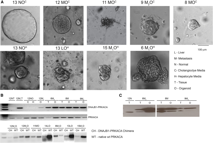
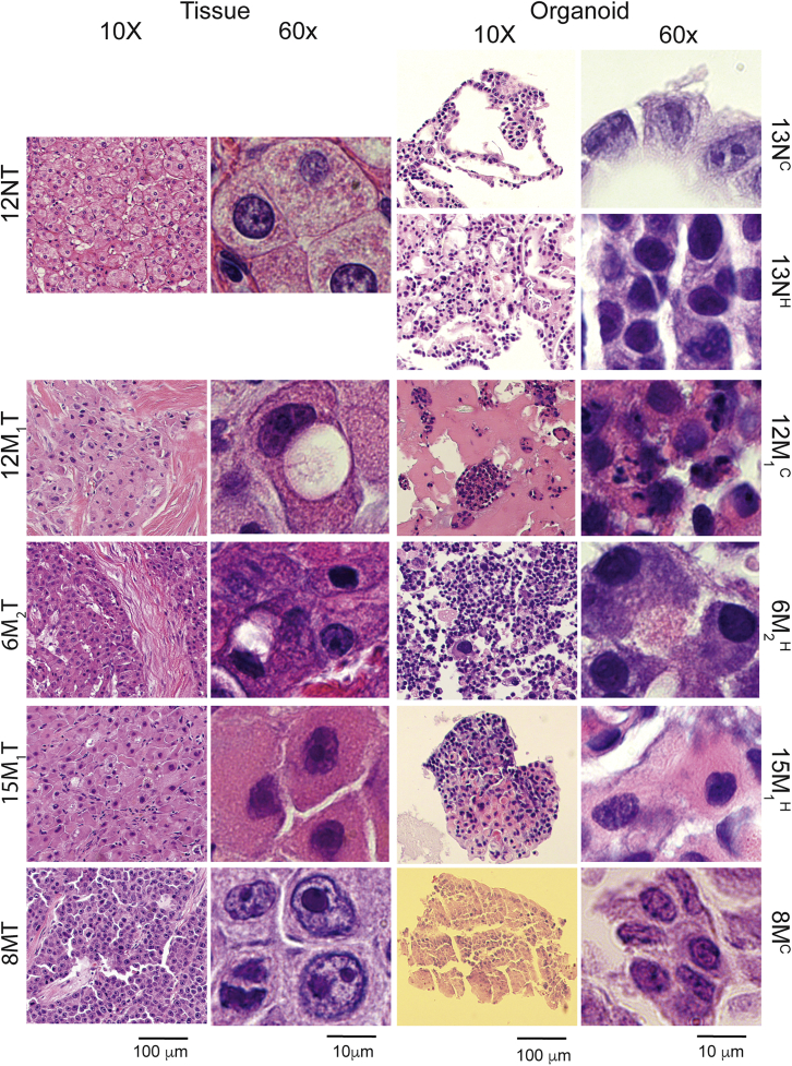
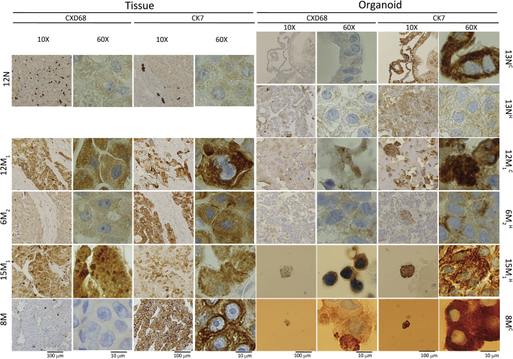
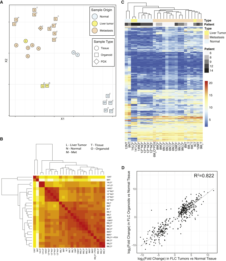
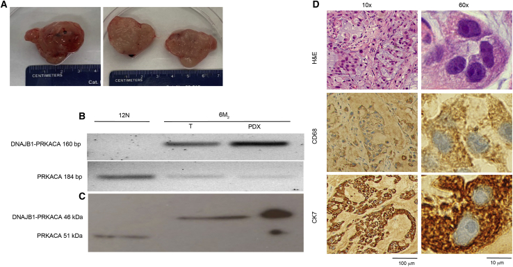
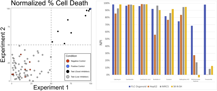
Fibrolamellar carcinoma (FLC) is a rare, often lethal, liver cancer affecting adolescents and young adults, for which there are no approved therapeutics. The development of therapeutics is hampered by a lack of in vitro models. Organoids have shown utility as a model system for studying many diseases. In this study, tumor tissue and the adjacent non-tumor liver were obtained at the time of surgery. The tissue was dissociated and grown as organoids. We developed 21 patient-derived organoid lines: 12 from metastases, three from the liver tumor and six from adjacent non-tumor liver. These patient-derived FLC organoids recapitulate the histologic morphology, immunohistochemistry, and transcriptome of the patient tumor. Patient-derived FLC organoids were used in a preliminary high-throughput drug screen to show proof of concept for the identification of therapeutics. This model system has the potential to improve our understanding of this rare cancer and holds significant promise for drug testing and development.

Keywords: fibrolamellar; liver cancer; pediatric cancer.


Figures

Figure 1
Figure 1
Brightfield imaging, RNA transcription, and protein expression of DNAJB1-PRKACA in patient-derived FLC organoids (A) Brightfield imaging of normal cholangiocyte and hepatocyte organoids compared with FLC primary liver tumor and metastatic organoids. Scale bar, 100 μM in all images. (B) The DNAJB1-PRKACA fusion transcript was detected in tumor tissue and tumor organoids but not in normal tissue or normal organoids. The wild-type PRKACA transcript was detected in all samples. Top pair of gels: DNAJB1-PRKACA amplicon = 160 bp, wild-type PRKACA amplicon = 184 bp. Bottom: DNAJB1-PRKACA amplicon = 104 bp, wild-type amplicon = 95 bp. (C) The DNAJB1-PRKACA fusion protein was detected in tumor tissue and tumor organoids but not in normal tissue from patients with FLC. DNAJB1-PRKACA protein = 46 kDa, wild-type PRKACA protein = 41 kDa. N, normal; M, M1, M2, metastasis #; P, primary tumor; O, organoid; T, tissue; C, cholangiocyte; H, hepatocyte.
Figure 2
Figure 2
Histology of FLC patient tumor tissue and patient-derived FLC organoids H&E-stained slides of normal cholangiocyte and hepatocyte organoids derived from normal tissue compared with FLC tumor organoids derived from their corresponding patient FLC tumor tissue. Slides were imaged at 10× and 60× magnification with scale bars of 100 and 10 μm, respectively. N, normal; M, M1, M2, metastasis #; C, cholangiocyte medium; H, hepatocyte medium; 13NC, passage two cholangiocyte medium; 13NH, passage six hepatocyte medium; 12M1, cholangiocyte medium; 6M2H, hepatocyte medium; 15M1H, hepatocyte medium.
Figure 3
Figure 3
Immunohistochemistry of patient-derived FLC organoids CD68- and CK7-stained slides of normal cholangiocyte and hepatocyte organoids derived from normal tissue compared with FLC tumor organoids derived from their corresponding patient FLC tumor tissue. Slides were imaged at 10× and 60× magnification with scale bars of 100 and 10 μm, respectively. N, normal; M, M1, M2, metastasis #. 13NC, passage two cholangiocyte medium; 13NH, passage six hepatocyte medium; 12M1C, cholangiocyte medium; 6M2, hepatocyte medium; 15M1, hepatocyte medium.
Figure 4
Figure 4
Transcriptome of patient-derived FLC organoids (A) Principal-component analysis comparing normal cholangiocyte and hepatocyte organoids derived from normal tissue compared with FLC tumor organoids (in cholangiocyte and hepatocyte media) derived from their corresponding patient FLC tumor tissue. (B) Heatmap organized by closest similarity between FLC patient normal tissue, FLC patient tumor tissue, patient-derived normal organoids, and FLC organoids in cholangiocyte or hepatocyte media. (C) Heatmap with known FLC-dysregulated genes comparing FLC patient normal tissue, FLC patient tumor tissue, patient-derived normal organoids, and FLC organoids in cholangiocyte or hepatocyte media. Both heatmaps show unsupervised clustering. N, normal; M, metastasis; L, primary liver tumor; O, organoid; T, tissue; H, grown in hepatocyte medium; C, grown in cholangiocyte medium. When multiple independent metastases were resected and analyzed, we used a subscript. Sample 14 was isolated in an alternative medium for liver tumors, which replaced the Noggin, Rspo-1, and Wnt3a with 3 nM dexamethasone (Broutier et al., 2017). (D) We have generated a “fibrolamellar signature” representing 509 transcripts (Lalazar et al., 2021). The change in expression of these genes in the FLC-derived organoids relative to organoids derived from non-transformed liver tissue was plotted as a function of the expression in FLC tumor relative to the adjacent non-transformed tissue. The Pearson correlation coefficient is > 0.8.
Figure 5
Figure 5
Patient-derived FLC organoids form tumors when injected into mice (A) Gross pathology of 6 M2 tumor after resection from mouse. On the left is the intact tumor and on the right a cross-section. (B) The DNAJB1-PRKACA fusion transcript (amplicon = 160 bp) was detected in original FLC patient tumor tissue (6M2 T) and tumor formed by injecting patient-derived organoids into mice (6 M2 O→M), but not in tissue from the adjacent non-transformed tissue (normal) from FLC patient (12 N T). The wild-type PRKACA transcript (amplicon = 184 bp) was detected in all samples. (C) The DNAJB1-PRKACA fusion protein (46 kDa) was detected in original FLC patient tumor tissue (6M2 T) and tumor formed by injecting patient-derived organoids into mice (6M2 PDX), but not in normal tissue from FLC patient (12N T). The wild-type PRKACA protein (41 kDa) was detected in all samples. (D) H&E-, CD68-, and CK7-stained slides of the tumor formed by injecting patient-derived organoids into mice (6M2 PDX). Scale bars, 100 μm at 10× magnification and 10 μm at 60× magnification. These organoids were grown in hepatocyte medium.
Figure 6
Figure 6
Preliminary drug screening of patient-derived FLC organoids (A) Correlation plots of screening ∼650 compounds against 6 M2 organoids performed twice (day 1 and day 2), each in singlet (R2 = 0.31; x axis, normalized %survival inhibition on day 1; y axis, normalized %survival inhibition on day 2. Blue dot, positive control (20 μM chaetocin); red dots, negative control (DMSO); gray dots, compounds tested. Compounds in the top right quandrant killed at least 50% 6 M2 organoids in both experiments. (B) Normalized percent inhibition of some of the most efficacious compounds against FLC organoids, HepG2 cells (from a 15-year-old Caucasian male who had a well-differentiated hepatocellular carcinoma), MRC5 (fibroblast cell line from lung), and SK-N-SH (neuroblastoma).

References

    1. Abdul-Al H.M., Wang G., Makhlouf H.R., Goodman Z.D. Fibrolamellar hepatocellular carcinoma: an immunohistochemical comparison with conventional hepatocellular carcinoma. Int. J. Surg. Pathol. 2010;18:313–318. doi: 10.1177/1066896910364229. - DOI - PubMed
    1. Broutier L., Mastrogiovanni G., Verstegen M.M., Francies H.E., Gavarro L.M., Bradshaw C.R., Allen G.E., Arnes-Benito R., Sidorova O., Gaspersz M.P., et al. Human primary liver cancer-derived organoid cultures for disease modeling and drug screening. Nat. Med. 2017;23:1424–1435. doi: 10.1038/nm.4438. - DOI - PMC - PubMed
    1. Calandrini C., Schutgens F., Oka R., Margaritis T., Candelli T., Mathijsen L., Ammerlaan C., van Ineveld R.L., Derakhshan S., de Haan S., et al. An organoid biobank for childhood kidney cancers that captures disease and tissue heterogeneity. Nat. Commun. 2020;11:1310. doi: 10.1038/s41467-020-15155-6. - DOI - PMC - PubMed
    1. Cattaneo C.M., Dijkstra K.K., Fanchi L.F., Kelderman S., Kaing S., van Rooij N., van den Brink S., Schumacher T.N., Voest E.E. Tumor organoid-T-cell coculture systems. Nat. Protoc. 2020;15:15–39. doi: 10.1038/s41596-019-0232-9. - DOI - PMC - PubMed
    1. Craig J.R., Peters R.L., Edmondson H.A., Omata M. Fibrolamellar carcinoma of the liver: a tumor of adolescents and young adults with distinctive clinico-pathologic features. Cancer. 1980;46:372–379. doi: 10.1002/1097-0142(19800715)46:2<372::AID-CNCR2820460227>3.0.CO;2-S. - DOI - PubMed

Publication types

Supplementary concepts

LinkOut - more resources