. 2022 Aug 10;30(8):1163-1172.e6.
doi: 10.1016/j.chom.2022.06.006. Epub 2022 Jul 7.

Non-protective immune imprint underlies failure of Staphylococcus aureus IsdB vaccine

Affiliations

Non-protective immune imprint underlies failure of Staphylococcus aureus IsdB vaccine

Chih-Ming Tsai et al. Cell Host Microbe. .

Abstract

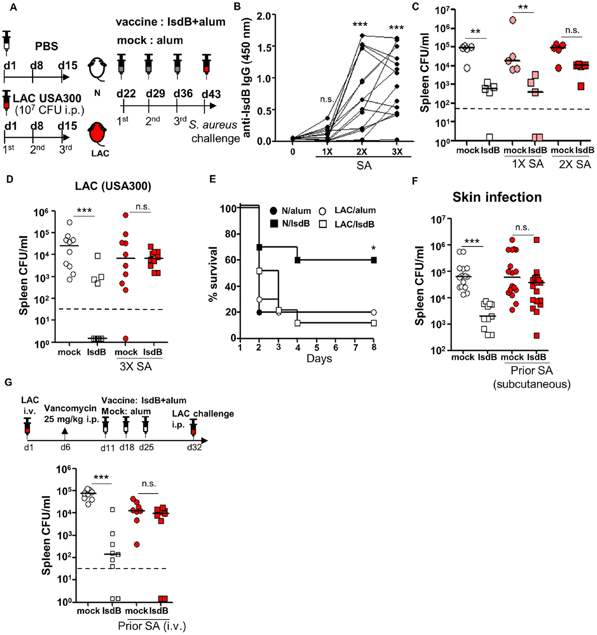
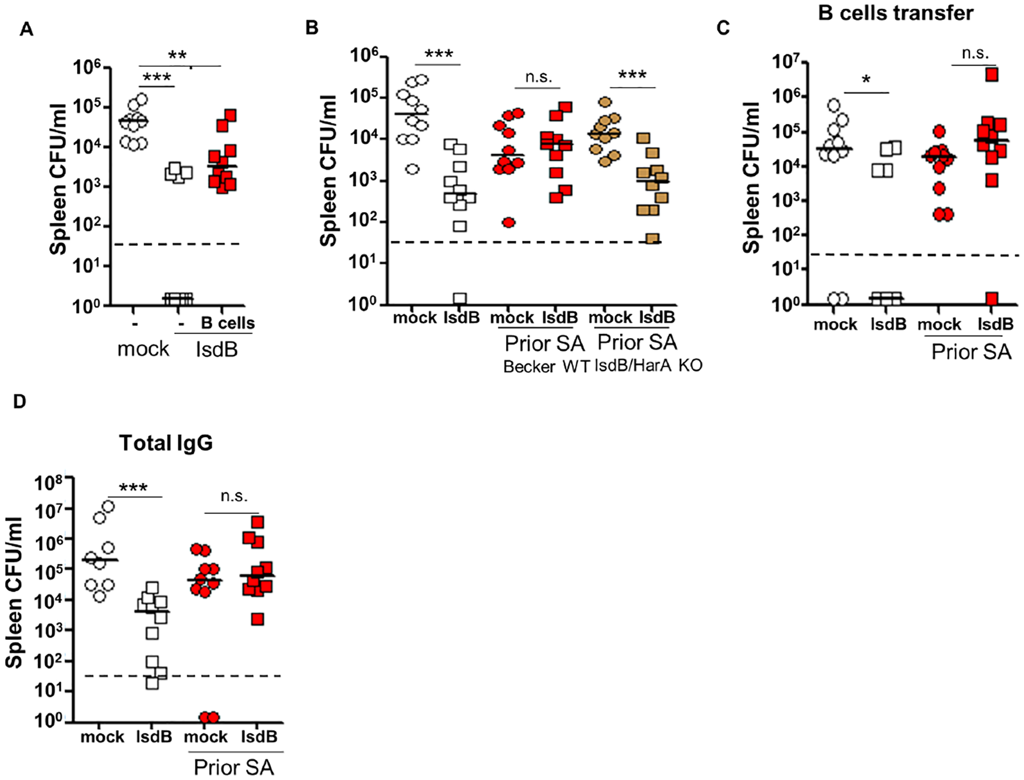
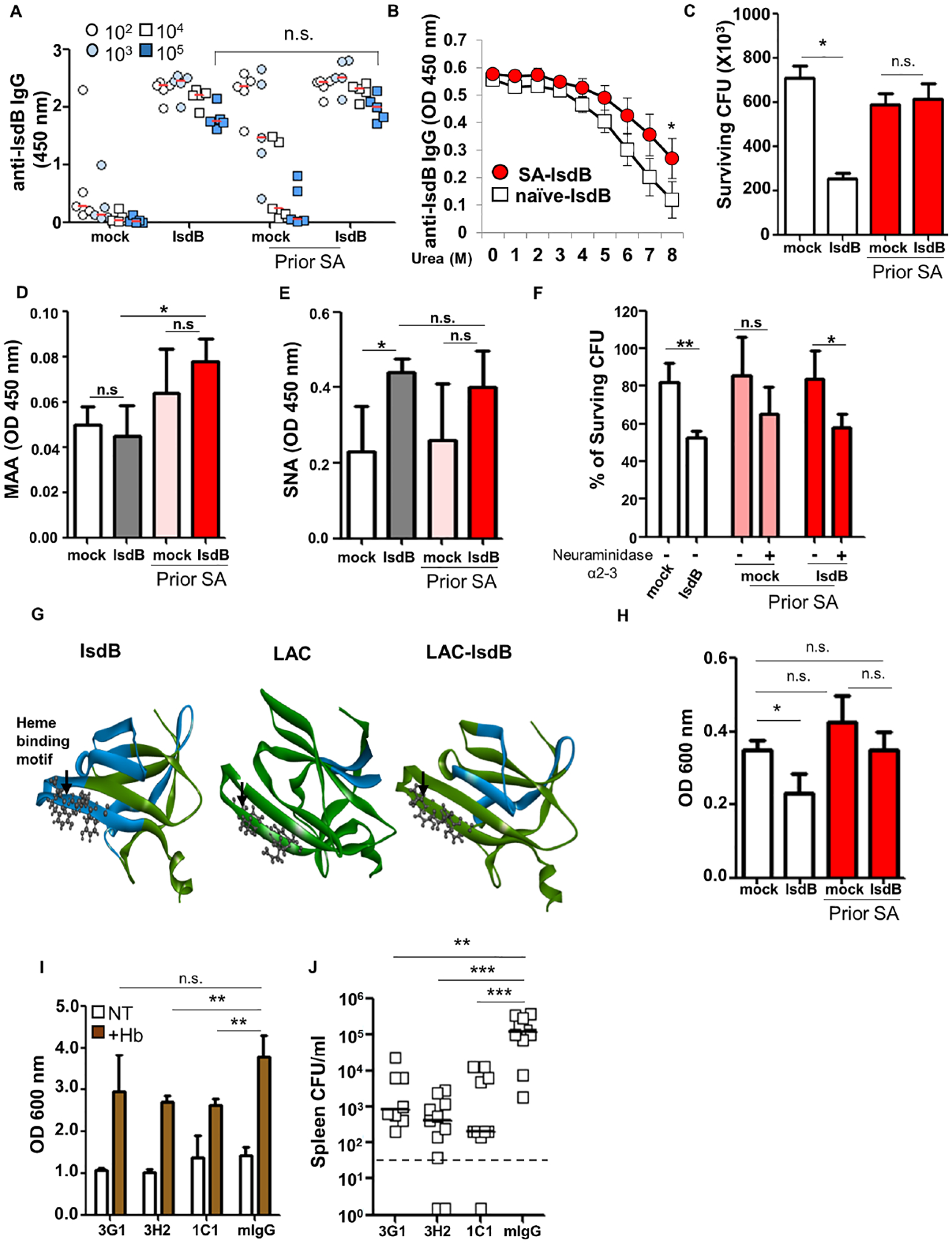
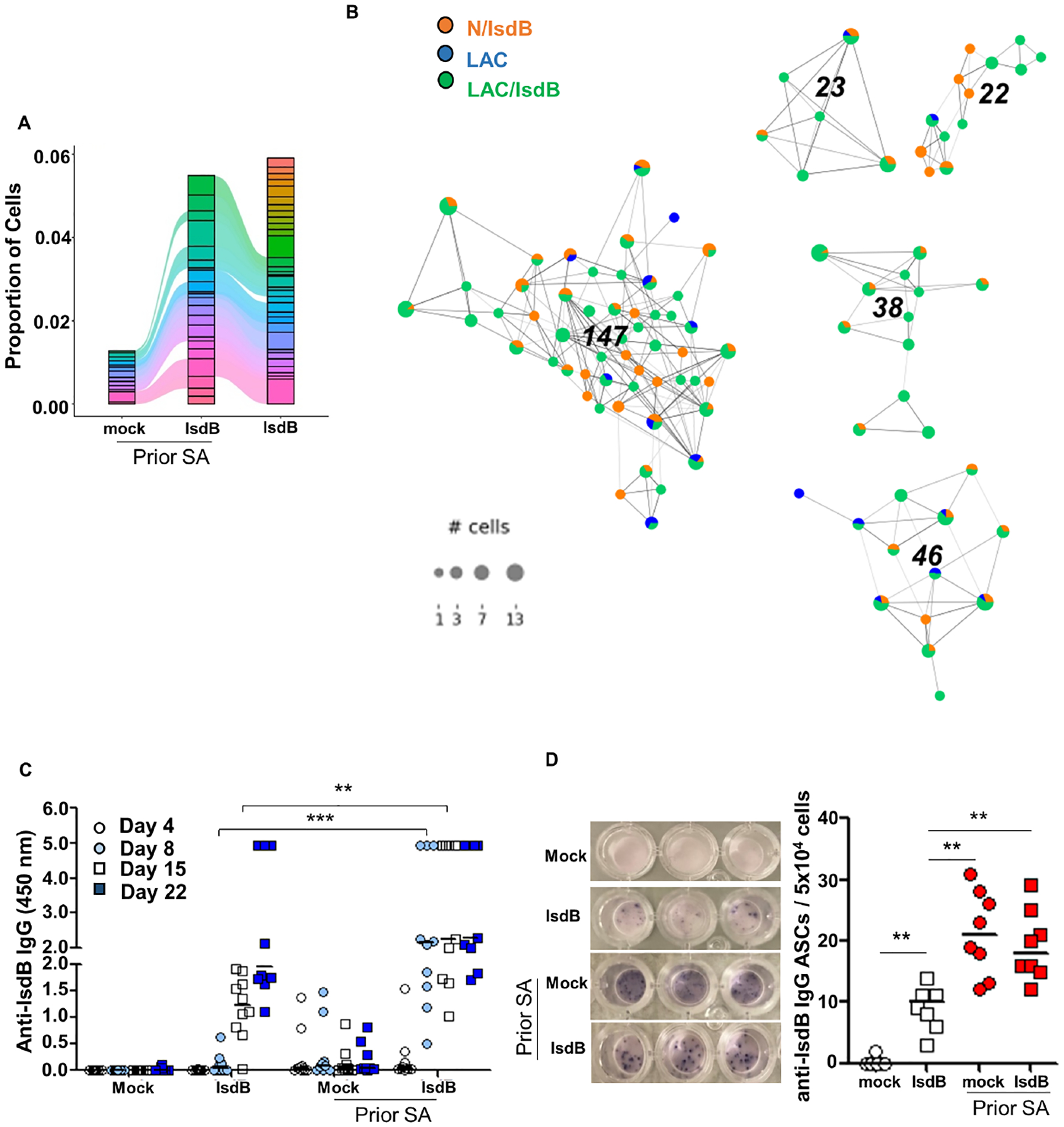
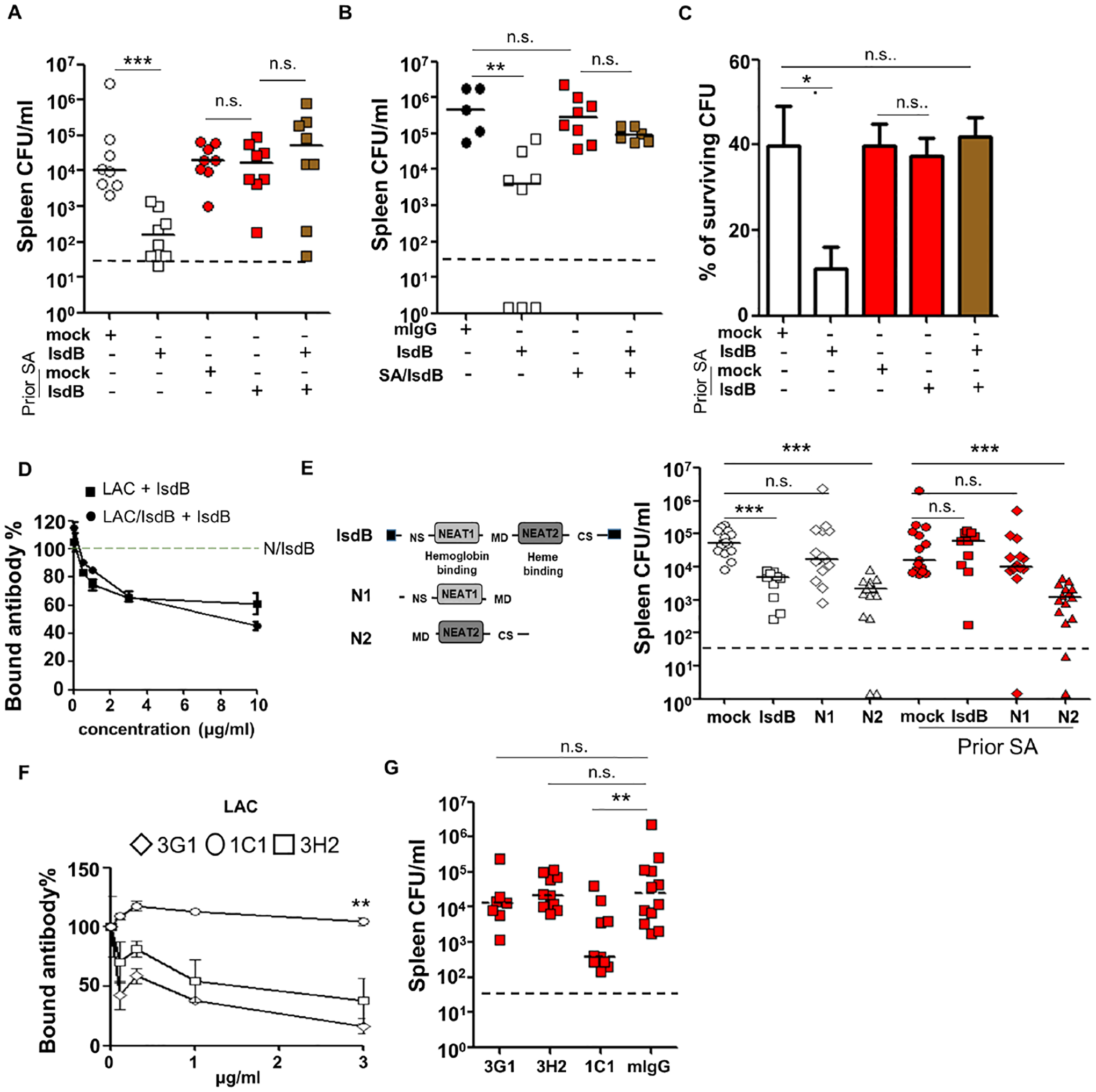
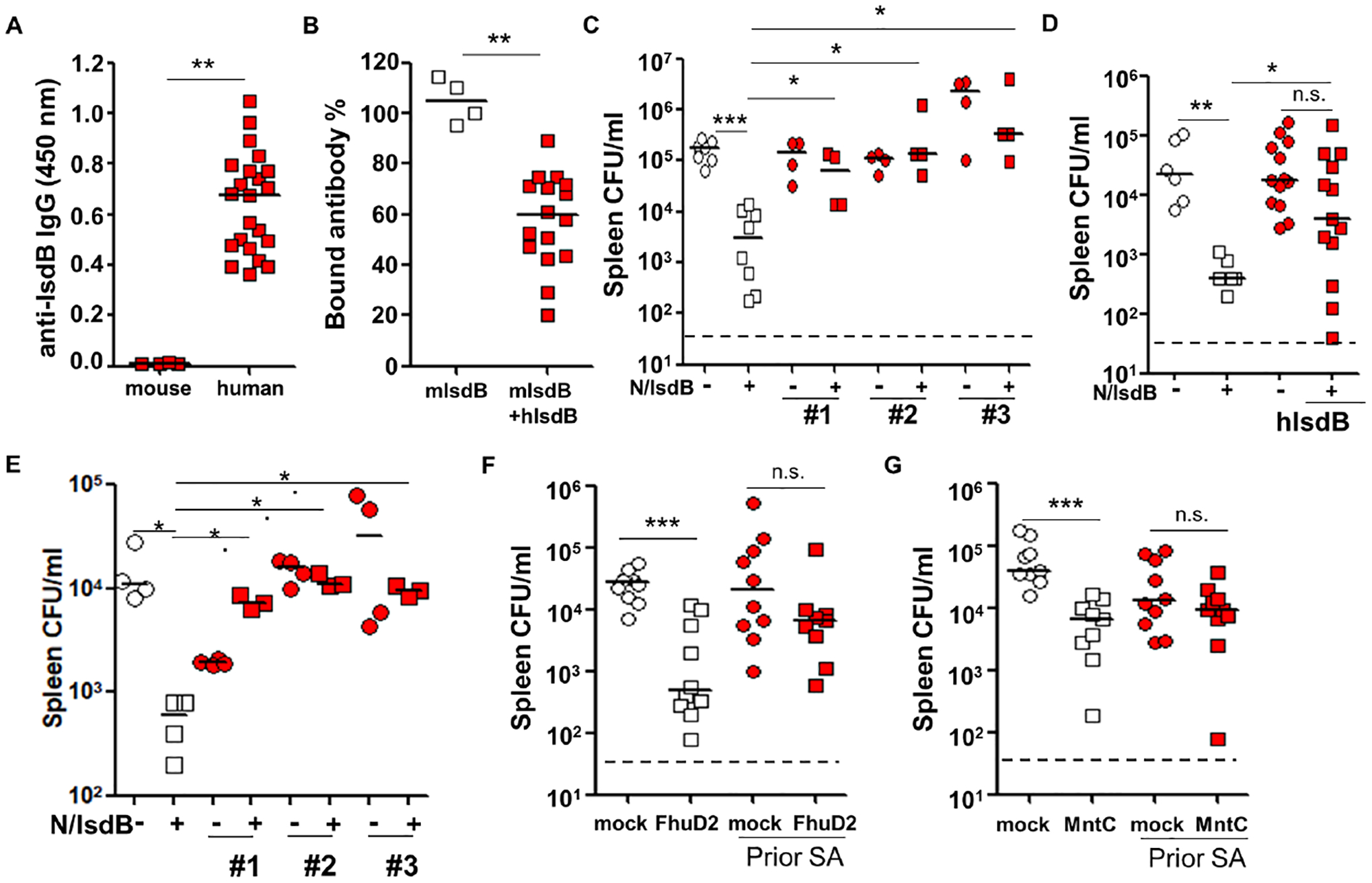
Humans frequently encounter Staphylococcus aureus (SA) throughout life. Animal studies have yielded SA candidate vaccines, yet all human SA vaccine trials have failed. One notable vaccine "failure" targeted IsdB, critical for host iron acquisition. We explored a fundamental difference between humans and laboratory animals-natural SA exposure. Recapitulating the failed phase III IsdB vaccine trial, mice previously infected with SA do not mount protective antibody responses to vaccination, unlike naive animals. Non-protective antibodies exhibit increased α2,3 sialylation that blunts opsonophagocytosis and preferentially targets a non-protective IsdB domain. IsdB vaccination of SA-infected mice recalls non-neutralizing humoral responses, further reducing vaccine efficacy through direct antibody competition. IsdB vaccine interference was overcome by immunization against the IsdB heme-binding domain. Purified human IsdB-specific antibodies also blunt IsdB passive immunization, and additional SA vaccines are susceptible to SA pre-exposure. Thus, failed anti-SA immunization trials could be explained by non-protective imprint from prior host-SA interaction.

Keywords: IsdB; S. aureus; Staphylococcus aureus; antibody; antibody competition; immunization; original antigenic sin; vaccine; vaccine failure.


Conflict of interest statement

Declaration of interests C.-M.T. and G.Y.L. have filed for a patent application for the use of IsdB NEAT2 as a vaccine.

Figures

Figure 1
Figure 1. IsdB immunization is not protective in mice previously infected with S. aureus
(A) Experimental setting. C57BL/6 mice injected intraperitoneally (i.p.) with 2×10⁷ SA (LAC) or phosphate-buffered saline (PBS) 3 times at 7d intervals, immunized i.p. with IsdB plus alum (IsdB) or alum alone (Mock), then challenged with 2×10⁷ LAC i.p. (B) Serum anti-IsdB IgG after 1–3 LAC infections (n=15 per condition). (C and D) Tissue bacterial burden in mice infected 1–3 times with LAC, then immunized and LAC challenged as per Figure 1A. Bacterial burden was measured 24 hrs after the last infection. N/IsdB: Naïve mice vaccinated with IsdB, N/alum: naïve mice given adjuvant alone; LAC/IsdB: LAC-infected mice vaccinated with IsdB; LAC/alum: LAC-infected mice given adjuvant alone (LAC/alum). N=5 for (C) and n=10 for (D) per mouse group. (E) Kaplan-Meyer plot of mice treated as in Figure 1A with a final LAC challenge dose of 5×10⁷ (LD90) (n=10 per mouse group). (F) Tissue bacterial burden in mice infected subcutaneously (twice 2 weeks apart) with SA (LAC), IsdB immunized two weeks later and then LAC challenged i.p. per Figure 1A (n=11–19 per mouse group). (G) Tissue bacterial burden in mice infected once i.v. with SA, treated for 5 days with vancomycin and then immunized and LAC challenged i.p. per Figure 1A (n=9 per mouse group). Each data point represents an individual mouse; bars denote median and dashed lines indicate the limit of detection (C, D, F and G). n.s., not significant, *P<0.05, **P<0.01, and ***P< 0.001; one-way ANOVA (B to D, F and G) or long rank test (E). See also Figure S1.
Figure 2
Figure 2. Parameters of IsdB vaccine and vaccine interference
(A) B cells adoptively transferred from SA (LAC) infected mice blunt IsdB vaccine efficacy in the recipient mice. Recipient mice were immunized with IsdB as per Figure 1A, 24h after B cell transfer (n=10 per mouse group). (B) Lack of vaccine interference when IsdB/HarA Becker strain is used for prior SA infection and WT Becker used in final challenge (n=10 per mouse group). (C and D) Prior LAC infection abrogates humoral protection conferred by IsdB vaccine. Naïve mice received splenic B cells (n=10 per mouse group) (C) or total IgG (mock, n=8; IsdB, LAC/mock and LAC/IsdB, n=10) (D) i.v. from donor mice infected and immunized as in Figure 1A. The recipient mice were infected 24h later with LAC, and tissue bacterial burden was measured after another 24 hrs. See also Figure S2. Each data point represents an individual mouse; bars denote median and dashed lines indicate the limit of detection. n.s., not significant, *P<0.05, **P<0.01, and ***P< 0.001; one-way ANOVA (A to D).
Figure 3
Figure 3. Differences in IsdB targets and sialylation contribute to functional differences of IsdB-specific antibodies
(A) Total serum anti-IsdB IgG on day 8 after IsdB immunization performed as per (Figure 1A) (n=5 per mouse group). (B) Avidity of IsdB-specific Ab measured in the presence of increasing urea concentrations. (n = 2). (C) Opsonophagocytic killing (OPK) of SA (LAC) by primary mouse neutrophils in the presence of immunized sera. The graph represents mean values ± SD from three independent experiments. (D and E) Sialylation of immunized sera IgG was determined by binding to Sambucus Nigra Lectin (SNA, α2–6 sialic acid specific) (H) and Maackia Amurensis Lectin II (MAA, α2–3 sialic acid specific) (I) in a lectin-ELISA. (F) OPK of SA (LAC) by primary mouse neutrophils in the presence of immunized sera with or without α2–3 neuraminidase for 16 hrs at 4 °C. (G) Ribbon representation of interaction between immunized mouse sera (from Figure 1A) and overlapping IsdB peptide library. Colors indicate Intensity of Ab binding to specific domains: green (0 or low intensity) to red (over 1000). White arrow points to heme-binding motif. (H) Heme-dependent growth of SA in the presence of immunized mouse sera. The graph represents mean values ± SD from three independent experiments. (I) Heme-dependent growth of SA in the presence of mAbs from clones derived from a IsdB-specific B cell hybridoma library, generated by IsdB vaccination of naïve mice. The graph represents mean values ± SD from three independent experiments. (J) Adoptive transfer of IsdB-specific mAbs from Figure 2E (0.5 mg/kg) prevents SA infection (n=8–11 per mouse group). Bar represents group median. Each data point represents an individual mouse (A and J); bars denote median and dashed lines indicate the limit of detection (J). Error bars represent means ± SD. n.s., not significant, *P<0.05, **P<0.01, and ***P< 0.001; One-way ANOVA (A to F, and H to J). See also Figure S3 and S4A.
Figure 4
Figure 4. IsdB vaccination recalls the non-protective IsdB-specific response from prior S. aureus infection
(A) Alluvial plot showing the top 40 of clonotypes from LAC, LAC/IsdB and N/IsdB. (B) IsdB-specific clonotype network. Each dot represents cells with identical CDR3 sequence. For each clonotype, the numeric clonotype ID is shown in the graph. The size of each dot refers to the number of cells with the same CDR3. (C) Rapid rise of specific IgG serum titer 7 days after first IsdB vaccination in SA pre-infected mice (n=10 per mouse group). (D) ELISpot assay showing IsdB-specific IgG-secreting cells (ASCs) per 5×10⁴ spleen B cells isolated 7 Days after the first IsdB vaccination. Error bars represent means ± SD. n.s., not significant, *P<0.05, **P<0.01, and ***P< 0.001; one-way ANOVA (C and D). See also Figure S4.
Figure 5
Figure 5. Competition for IsdB between non-protective and protective antibodies determines outcome of S. aureus infection and overcoming antibody competition
(A) Anti-SA (LAC) immunity of mice co-injected with equal volume (50 μl i.p.) of protective (NT-IsdB) and non-protective (LAC-IsdB) IsdB-specific sera (n=8 per mouse group). (B) Anti-SA (LAC) immunity of mice co-injected with equal amount (25 μg i.p.) of protective (NT-IsdB) and non-protective (LAC-IsdB) IsdB-specific Ab (n=5–8 per mouse group). (C) Effect of IsdB-specific Ab competition on mouse neutrophil OPK. Graph represents mean values ± SD from three independent experiments. (D) IsdB binding to biotinylated IsdB-specific Ab from NT/IsdB (1 μg/ml) in the presence of IsdB-specific Ab from LAC or LAC/IsdB immunized mice in an ELISA plate assay. (E) Schematic of the IsdB, N1 and N2 proteins used for vaccination. Efficacy of N1 and N2 immunization in naïve and LAC pre-infected mice (n=10–15 per mouse group). (F) IsdB binding by biotinylated mAbs (0.5 μg/ml) in the absence or presence of IsdB-specific Ab purified from LAC-infected mice, in an ELISA plate assay. (G) Efficacy of adoptively transferred anti-IsdB mAbs (0.5 mg/kg i.p.) in preventing SA-infection in naïve and SA (LAC) pre-infected mice (n=7–10 per mouse group). Each data point represents an individual mouse, bars denote median and dashed lines indicate the limit of detection (A, B, E and G). Error bars represent means ± SD. n.s., not significant, *P<0.05, **P<0.01, and ***P< 0.001; Student’s t test (C and D) or one way ANOVA (A, B and E to G). See also Figure S5.
Figure 6
Figure 6. Human serum anti-staphylococcal antibodies blunting of protection conferred by anti-IsdB passive immunization, and interference with FhuD2 and MntC vaccines
(A) Total anti-IsdB IgG titer in human adult and naïve laboratory mouse sera, diluted to 1:10,000 (mouse sera, n=4; human sera, n=22). (B) IsdB binding by biotinylated IsdB-specific mouse Ab (1 μg) generated by vaccine in the absence or presence of IsdB-specific human Ab (10 μg) in an ELISA plate assay (human sera, n=16). (C) Anti-SA protection conferred by co-transfer of IsdB-vaccinated mouse sera (50 μl i.v.) and serum from 3 human donors (100 μl i.p.) (n=4–8 per mouse group). (C) Anti-SA protection conferred by sequential transfer of purified human IsdB-specific Ab (or control IgG, time = 0 hr) and vaccine-generated IsdB-Ab in mice (time = 16 hr). Ratio of protective to non-protective Ab injected IV: 25 μg to 35 μg. Each red square or circle represents mean SA burden from 3–5 mice (human sera, n=13). (D) Anti-SA protection conferred by sequential transfer of purified human IsdB-specific Ab (or control IgG, time = 0 hr) and vaccine-generated IsdB-Ab in mice (time = 16 hr). Ratio of protective to non-protective Ab injected IV: 25 μg to 2.5 μg (human Ab, n = 3). (F and G) Tissue bacterial burden in mice infected 3 times with LAC, then immunized with FhuD2 (F) or MntC (G) and LAC challenged as per Fig. 1A. Bacterial burden is measured 24 hrs after LAC challenge. Each data point represents an individual mouse serum or human serum (A, B and D). Bars denote median and dashed lines indicate the limit of detection (C, D, F and G). Error bars represent means ± SD. n.s., not significant, *P<0.05, **P<0.01, and ***P< 0.001; Student’s t test (A and B) or one way ANOVA (C to G). See also Figure S6.

References

    1. Alonzo F 3rd, Kozhaya L, Rawlings SA, Reyes-Robles T, DuMont AL, Myszka DG, Landau NR, Unutmaz D, and Torres VJ (2013). CCR5 is a receptor for Staphylococcus aureus leukotoxin ED. Nature 493, 51–55. - PMC - PubMed
    1. Anderson AS, Scully IL, Timofeyeva Y, Murphy E, McNeil LK, Mininni T, Nunez L, Carriere M, Singer C, Dilts DA, et al. (2012). Staphylococcus aureus manganese transport protein C is a highly conserved cell surface protein that elicits protective immunity against S. aureus and Staphylococcus epidermidis. J Infect Dis 205, 1688–1696. - PMC - PubMed
    1. Bennett MR, Dong J, Bombardi RG, Soto C, Parrington HM, Nargi RS, Schoeder CT, Nagel MB, Schey KL, Meiler J, et al. (2019). Human VH1–69 Gene-Encoded Human Monoclonal Antibodies against Staphylococcus aureus IsdB Use at Least Three Distinct Modes of Binding To Inhibit Bacterial Growth and Pathogenesis. mBio 10. - PMC - PubMed
    1. Borcherding N, Bormann NL, and Kraus G (2020). scRepertoire: An R-based toolkit for single-cell immune receptor analysis. F1000Res 9, 47. - PMC - PubMed
    1. Daily J (2016). Parasail: SIMD C library for global, semi-global, and local pairwise sequence alignments. BMC Bioinformatics 17, 81. - PMC - PubMed