. 2022 Jul 8;13(1):3971.
doi: 10.1038/s41467-022-31663-z.

The cholesterol uptake regulator PCSK9 promotes and is a therapeutic target in APC/KRAS-mutant colorectal cancer

Affiliations

The cholesterol uptake regulator PCSK9 promotes and is a therapeutic target in APC/KRAS-mutant colorectal cancer

Chi Chun Wong et al. Nat Commun. .

Abstract

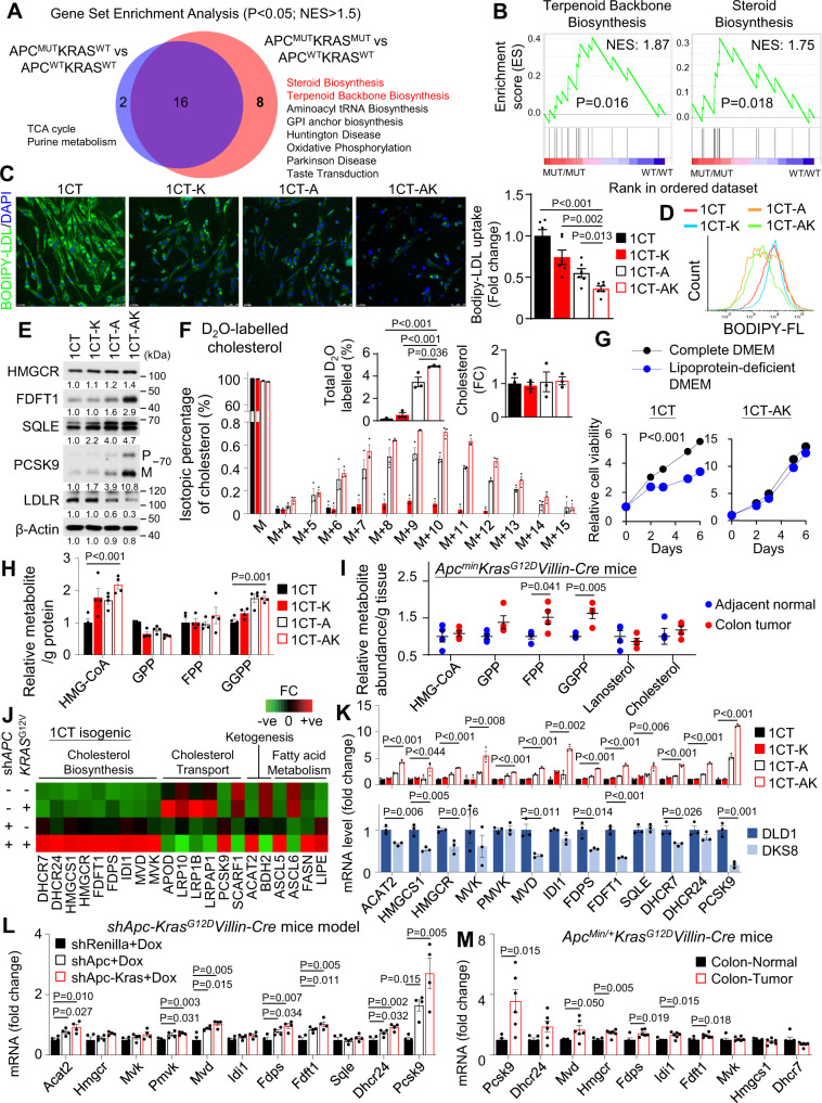
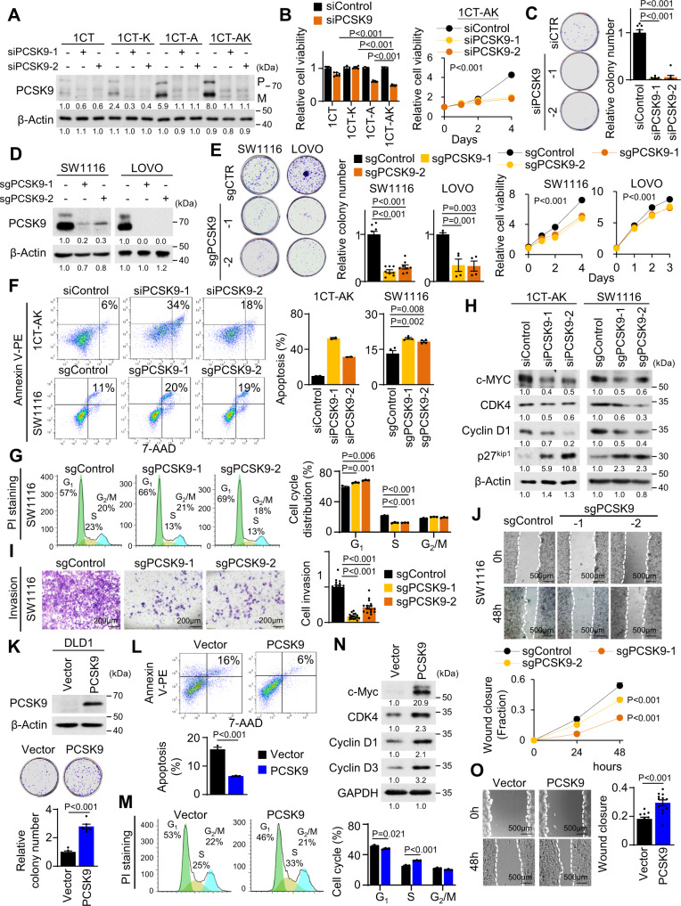
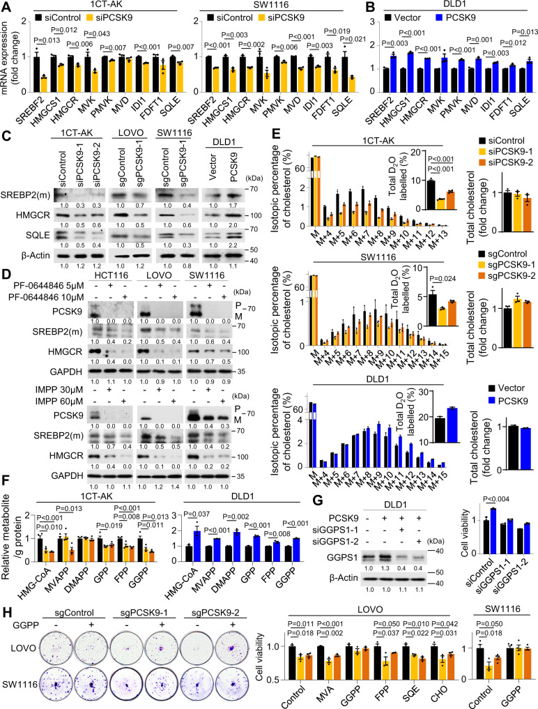
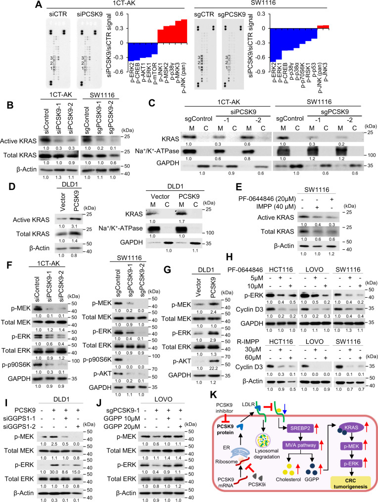
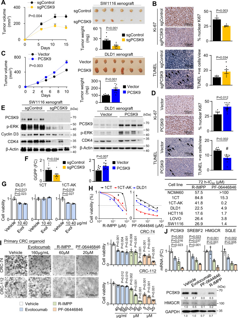
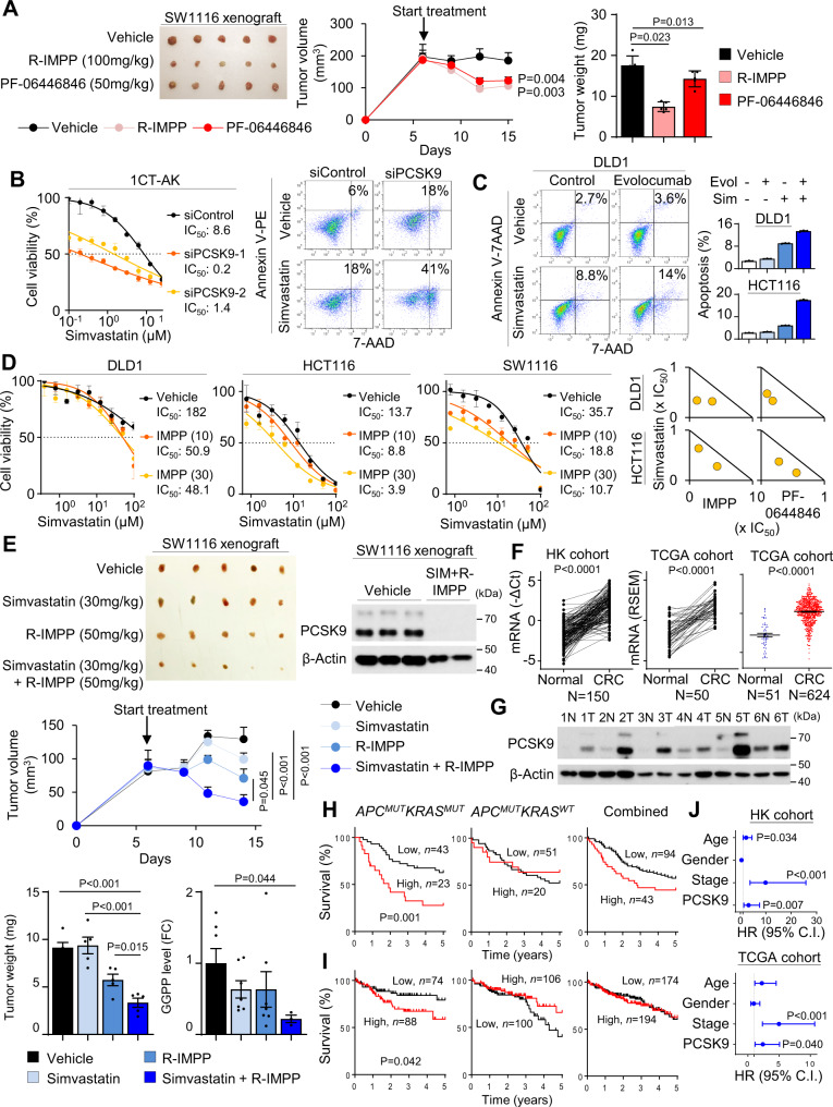
Therapeutic targeting of KRAS-mutant colorectal cancer (CRC) is an unmet need. Here, we show that Proprotein Convertase Subtilisin/Kexin type 9 (PSCK9) promotes APC/KRAS-mutant CRC and is a therapeutic target. Using CRC patient cohorts, isogenic cell lines and transgenic mice, we identify that de novo cholesterol biosynthesis is induced in APC/KRAS mutant CRC, accompanied by increased geranylgeranyl diphosphate (GGPP)─a metabolite necessary for KRAS activation. PCSK9 is the top up-regulated cholesterol-related gene. PCSK9 depletion represses APC/KRAS-mutant CRC cell growth in vitro and in vivo, whereas PCSK9 overexpression induces oncogenesis. Mechanistically, PCSK9 reduces cholesterol uptake but induces cholesterol de novo biosynthesis and GGPP accumulation. GGPP is a pivotal metabolite downstream of PCSK9 by activating KRAS/MEK/ERK signaling. PCSK9 inhibitors suppress growth of APC/KRAS-mutant CRC cells, organoids and xenografts, especially in combination with simvastatin. PCSK9 overexpression predicts poor survival of APC/KRAS-mutant CRC patients. Together, cholesterol homeostasis regulator PCSK9 promotes APC/KRAS-mutant CRC via GGPP-KRAS/MEK/ERK axis and is a therapeutic target.


Conflict of interest statement

The authors declared no competing interests.

Figures

Fig. 1
Fig. 1. Cholesterol homeostasis is shifted from cellular uptake to de novo biosynthesis in APC/KRAS-mutant CRC leading to increase geranylgeranyl pyrophosphate (GGPP) levels.
A Gene Set Enrichment Analysis (GSEA) of enriched KEGG pathways in APC-mutant (N = 142), APC/KRAS-mutant CRC (N = 134) as compared to wildtype counterparts (N = 50) from the Cancer Genome Atlas (TCGA) colon and rectal cancer (COADREAD) cohort. Permutation-based p-value. B Terpenoid Backbone Biosynthesis pathway and Steroid Biosynthesis pathway are enriched in APC/KRAS-mutant CRC cases from TCGA cohort. Permutation based p-value. C Fluorescence microscopy of cellular uptake of fluorescent labelled LDL (BODIPY-LDL) in 1CT isogenic cell lines (1CT-K: 1CT-KRASG12V, 1CT-A: 1CT-shAPC, 1CT-AK: 1CT-shAPC-KRASG12V). Reduced LDL uptake was observed in 1CT-AK cells (n = 6). D Flow cytometry of intracellular fluorescence after incubation with BODIPY-LDL. E Western blot confirmed the up-regulated protein expression of PCSK9 and cholesterol biosynthesis genes in 1CT-AK cells; LDLR was inhibited. F D2O stable isotope labeling (48 h) of cholesterol and LC-MS analysis of intracellular cholesterol in 1CT isogenic cells, revealing increased de novo cholesterol biosynthesis in 1CT-A and 1CT-AK cells, while having no effect of total cholesterol level (n = 3). G Cell viability of 1CT-AK cells were not sensitive to lipoprotein depletion in culture medium. Cell growth curve of 1CT and 1CT-AK cells in complete or lipoprotein-depleted medium (n = 3). H LC-MS of cholesterol pathway intermediates in 1CT isogenic cells. I LC-MS of cholesterol pathway intermediates in CRC and adjacent normal tissues from 7-weeks-old Apcmin/+KrasG12D/+Villin-Cre mice (n = 4). J PCR array (Human lipoprotein signaling & cholesterol metabolism; and Human fatty acid metabolism) revealed that KRASG12V plus shAPC increased mRNA expression of cholesterol biosynthesis genes. K qPCR validated the increased mRNA expression of PCSK9 and cholesterol biosynthesis genes in 1CT-AK cells compared to 1CT, 1CT-K, and 1CT-A cells, whilst knockout of mutant KRAS in DLD1 cells (i.e., DKS8) exerted an opposite effect (n = 3). L PCSK9 and cholesterol biosynthesis genes were up-regulated in the shApc-KrasG12D-Villin-Cre mice colon (n = 4) compared to shRen controls (n = 4) and shApc-Villin-Cre mice (n = 4). M mRNA levels of PCSK9 and cholesterol biosynthesis genes in ApcMin/+KrasG12DVillin-Cre mice tumors (n = 6) compared to adjacent normal tissues (n = 5). Data shown are means of biological replicates; ± SEM (C, FI, KM). Two-tailed Student’s t-test (C, F, H, I, KM). Two-tailed two-way ANOVA (G). Source data are provided as a Source Data file.
Fig. 2
Fig. 2. PCSK9 promotes malignant phenotypes in APC/KRAS-mutant CRC.
A PCSK9 knockdown in 1CT isogenic cell lines. B PCSK9 knockdown suppressed cell proliferation (n = 6) and C colony formation (n = 8) in 1CT-AK cells. D PCSK9 knockout in SW1116 and LOVO cells. E PCSK9 knockout reduced cell growth (n = 6) and colony formation (SW1116: n = 8, 7 days; LOVO: n = 4, 14 days) in SW1116 and LOVO cells. F PCSK9 knockdown (72 h) or knockout induced cell apoptosis (1CT-AK: n = 2; SW1116: n = 4) and G inhibited G1-S cell cycle progression (n = 3). H Western blot revealed that PCSK9 knockdown/knockout induced the expression of cleaved PARP and p27Kip1, while suppressing c-Myc, CDK4 and Cyclin D1. I PCSK9 knockout suppressed Matrigel invasion (72 h, 16 replicates in 4 independent experiments) and J wound healing closure (12 replicates in 4 independent experiments) in SW1116 cells. K PCSK9 overexpression in DLD1 cells increased cell proliferation (n = 6, 7 days), L inhibited apoptosis (n = 3) and M promoted G1-S progression (n = 3). N PCSK9 increased CDK4, Cyclin D1, and Cyclin D3, but suppressed p27Kip1 in DLD1 cells. O PCSK9 overexpression promoted cell migration in DLD1 cells (48 h, 12 replicates in 4 independent experiments). Data shown are means of biological replicates; ± SEM (B, C, EG, IM, O). Two-tailed Student’s t-test (B, C, EG, I, KM, O). Two-tailed two-way ANOVA (growth curves in B, E, J). Source data are provided as a Source Data file.
Fig. 3
Fig. 3. PCSK9 activates de novo cholesterol biosynthesis and GGPP accumulation to promote cell growth.
A mRNA expression of SREBF2 and cholesterol biosynthesis genes was decreased in PCSK9 knockdown 1CT-AK cells and PCSK9 knockout SW1116 cells (n = 3). B Ectopic expression of PCSK9 in DLD1 cells promoted SREBP2 and cholesterol biosynthesis genes mRNA expression (n = 3). C Western blot showed that PCSK9 knockdown or knockout reduced protein expression of SERBP2 (mature form), HMGCR, and SQLE in 1CT-AK, LOVO, and SW1116 cells; PCSK9 overexpression in DLD1 cells exerted an opposite effect. D R-IMPP and PF-0644846, inhibitors of PCSK9, suppressed expression of SREBP2, HMGCR, and SQLE in APC/KRAS-mutant CRC cell lines. E D2O-labeling (48 h) and LC-MS analysis of intracellular cholesterol demonstrated that PCSK9 depletion inhibited de novo cholesterol biosynthesis in 1CT-AK (n = 4) and SW1116 cells (n = 3), while PCSK9 overexpression promoted de novo cholesterol biosynthesis in DLD1 cells (n = 2). F PCSK9 depletion reduced HMG-CoA, GPP, FPP and GGPP levels in 1CT-AK cells (n = 4), while PCSK9 overexpression increased GPP, FPP and GGPP in DLD1 cells (n = 4). G GGPS1 knockdown (n = 3, 72 h) abrogated growth inducing effect of PCSK9 overexpression in DLD1 cells. H Effect of cholesterol and intermediary metabolites in rescuing the proliferation of PCSK9 knockout SW1116 (n = 4) and LOVO (n = 3) cells. Data shown are means of biological replicates; ± SEM (A, B, EH). Two-tailed Student’s t-test (A, B, EH). Source data are provided as a Source Data file.
Fig. 4
Fig. 4. PCSK9 promotes KRAS/MEK/ERK signaling cascade via GGPP.
A Phospho-MAPK antibody array assay of 1CT-AK and SW1116 cells with PCSK9 knockdown or knockout, respectively. p-ERK1/2 and p-CREB were consistently down-regulated. B Active KRAS pulldown assays revealed that PCSK9 depletion suppressed KRAS activation. C Loss of PCSK9 decreased the membrane localization of KRAS. D PCSK9 overexpression in DLD1 cells increased active KRAS expression and its membrane localization. E PCSK9 inhibitors, R-IMPP and PF-0644846, suppressed the activation of KRAS in 1CT-AK and SW1116 cells. F PCSK9 knockdown/knockout in 1CT-AK and SW1116 cells inhibited phosphorylation of MEK, ERK, and p09S6K. G PCSK9 overexpression induced p-MEK and p-ERK in DLD1 cells. H R-IMPP and PF-0644846 inhibited p-ERK and expression of its downstream target Cyclin D3 in APC/KRAS-mutant CRC cell lines. I GGPS1 knockdown abrogated the induction of p-MEK and p-ERK by PCSK9 overexpression in DLD1 cells. J Supplementation of GGPP rescued p-MEK and p-ERK expression in PCSK9 knockout LOVO cells. K Schematic diagram summarizing the molecular mechanism of PCSK9 in APC/KRAS-mutant CRC. Source data are provided as a Source Data file.
Fig. 5
Fig. 5. PCSK9 promotes APC/KRAS-mutant CRC growth in vivo and growth inhibitory effect of PCSK9 inhibitors.
A PCSK9 knockout suppressed growth of subcutaneous xenograft model of SW1116 cells in NOD/SCID mice (n = 1 experiment, n = 9 mice per group). B PCSK9 knockout inhibited cell growth as indicated by Ki-67 staining (n = 5); TUNEL showed that PCSK9 knockout induced apoptosis in SW1116 xenografts (n = 5). C PCSK9 overexpression in DLD1 cells induced xenograft growth in nude mice (n = 1 experiment, n = 10 mice per group). D PCSK9 overexpression in DLD1 cells induced cell growth and inhibited apoptosis, as evidenced by Ki-67 (n = 7) and TUNEL (n = 8) assays, respectively. E Western blot of SW1116 xenografts revealed that PCSK9 knockout suppressed p-ERK, Cyclin D1, and CDK4. Western blot of DLD1 xenografts demonstrated increased expression of p-ERK, Cyclin D1, and CDK4. F GGPP levels in SW116 xenografts with PCSK9 knockout (n = 8) and DLD1 xenografts with PCSK9 overexpression (n = 10). G Effect of Evolocumab on 1CT, 1CT-AK, and DLD1 cells (n = 3). H Effect of R-IMPP and PF-0644846 on 1CT, 1CT-AK, and DLD1 cells (n = 3). 72 h-IC50 values of R-IMPP and PF-0644846 in a panel of APC/KRAS mutant CRC, 1CT isogenic cells and NCM460 cells. I Effect of Evolocumab, R-IMPP, and PF-0644846 on cell viability of primary organoids from human APC/ KRAS-mutant CRC after 5–7 days of treatment (n = 4). J Effect of Evolocumab, R-IMPP and PF-0644846 on cholesterol gene expression in KRAS-mutant CRC organoids (n = 3) after 72 h of drug treatment. Data shown are means of biological replicates; ± SEM (AD, F, GJ). Two-tailed Student’s t-test (AD, F, G, I, J). Two-tailed two-way ANOVA for growth curves (a, c). Source data are provided as a Source Data file.
Fig. 6
Fig. 6. PCSK9 and simvastatin synergistically inhibited APC/KRAS-mutant CRC growth and clinical significance of PCSK9 in CRC.
A Nude mice harboring SW1116 xenografts were treated with vehicle (PBS), R-IMPP (100 mg/kg), or PF-06446846 (50 mg/kg) (n = 1 experiment, n = 5 mice per group) by intraperitoneal injection every 2 days. Both R-IMPP and PF-06446846 inhibited tumor growth. B siPCSK9 synergized with simvastatin to inhibit cell growth and induce apoptosis of 1CT-AK cells (n = 3, 72 h). C Evolocumab synergized with simvastatin to promote cell apoptosis in KRAS-mutant CRC cells (96 h). D The co-administration of R-IMPP or PF-0644846 reduced 48 h-IC50 of simvastatin in KRAS-mutant CRC cells (left) (n = 3). Isobologram analyses indicated that combination of R-IMPP or PF-0644846 plus simvastatin were synergistic in suppressing cell viability (right). E Nude mice harboring SW1116 xenografts were treated with vehicle, R-IMPP, simvastatin, or their combination (n = 1 experiment, n = 5 mice per group). The combination drug treatment induced tumor regression and significantly suppressed tumor weight, PCSK9 protein expression and GGPP levels compared to vehicle or single treatment. F PCSK9 mRNA is up-regulated in CRC tissues as compared with paired adjacent normal tissues in Hong Kong (n = 150 pairs), and TCGA cohorts (n = 50 pairs), and in unpaired samples from TCGA cohort (n = 675). G PCSK9 protein expression is increased in CRC compared to adjacent normal tissues assessed by western blot. H Tissue microarray cohort (n = 137) showed that PCSK9 protein expression predicts poor survival of APCMUTKRASMUT CRC (n = 66), but not in APCMUTKRASWT CRC (n = 71) or overall cohort. Log-rank test (two-tailed). I PCSK9 mRNA predicts poor patient survival in APCMUTKRASMUT CRC (n = 162) in TCGA cohort, but not in APCMUTKRASWT CRC (n = 206) or overall cohort (n = 368). Log-rank test (two-tailed). J Multivariate COX proportional hazards regression analysis of the prognostic value of PCSK9 in APCMUTKRASMUT CRC patients in both Hong Kong and TCGA cohorts (n = 162, biological replicates). COX proportional hazards regression analysis. Data shown are means of biological replicates; ± SEM (AF). Two-tailed Student’s t-test (A, E, F). Two-tailed two-way ANOVA for growth curves (A, E). Source data are provided as a Source Data file.

References

    1. Siegel RL, Miller KD, Jemal A. Cancer statistics, 2020. CA Cancer J. Clin. 2020;70:7–30. doi: 10.3322/caac.21590. - DOI - PubMed
    1. Cancer Genome Atlas Network. Comprehensive molecular characterization of human colon and rectal cancer. Nature. 2013;487:330–337. doi: 10.1038/nature11252. - DOI - PMC - PubMed
    1. Janssen KP, et al. APC and oncogenic KRAS are synergistic in enhancing Wnt signaling in intestinal tumor formation and progression. Gastroenterology. 2006;131:1096–1109. doi: 10.1053/j.gastro.2006.08.011. - DOI - PubMed
    1. Roock WD, et al. KRAS, BRAF, PIK3CA, and PTEN mutations: Implications for targeted therapies in metastatic colorectal cancer. Lancet Oncol. 2011;12:594–603. doi: 10.1016/S1470-2045(10)70209-6. - DOI - PubMed
    1. Kuzu OF, Noory MA, Robertson GP. The role of cholesterol in cancer. Cancer Res. 2016;76:2063–2070. doi: 10.1158/0008-5472.CAN-15-2613. - DOI - PMC - PubMed

Publication types

MeSH terms