. 2022 Jul 8;12(1):11606.
doi: 10.1038/s41598-022-15809-z.

Polymicrobial biofilms of ocular bacteria and fungi on ex vivo human corneas

Affiliations

Polymicrobial biofilms of ocular bacteria and fungi on ex vivo human corneas

Konduri Ranjith et al. Sci Rep. .

Abstract

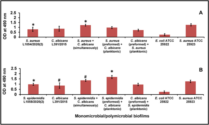
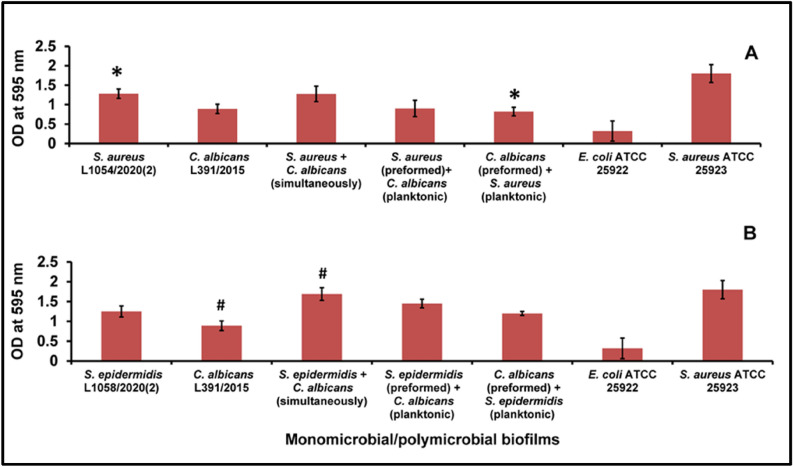
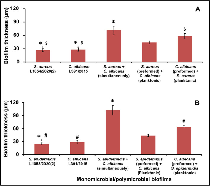
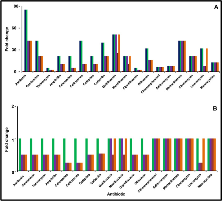
Microbes residing in biofilms confer several fold higher antimicrobial resistances than their planktonic counterparts. Compared to monomicrobial biofilms, polymicrobial biofilms involving multiple bacteria, multiple fungi or both are more dominant in nature. Paradoxically, polymicrobial biofilms are less studied. In this study, ocular isolates of Staphylococcus aureus, S. epidermidis and Candida albicans, the etiological agents of several ocular infections, were used to demonstrate their potential to form mono- and polymicrobial biofilms both in vitro and on human cadaveric corneas. Quantitative (crystal violet and XTT methods) and qualitative (confocal and scanning electron microscopy) methods demonstrated that they form polymicrobial biofilms. The extent of biofilm formation was dependent on whether bacteria and fungi were incubated simultaneously or added to a preformed biofilm. Additionally, the polymicrobial biofilms exhibited increased resistance to different antimicrobials compared to planktonic cells. When the MBECs of different antibacterial and antifungal agents were monitored it was observed that the MBECs in the polymicrobial biofilms was either identical or decreased compared to the monomicrobial biofilms. The results are relevant in planning treatment strategies for the eye. This study demonstrates that ocular bacteria and fungi form polymicrobial biofilms and exhibit increase in antimicrobial resistance compared to the planktonic cells.


Conflict of interest statement

The authors declare no competing interests.

Figures

Figure 1
Figure 1
Quantification of polymicrobial biofilm formation in S. aureus (A) and S. epidermidis (B) with C. albicans by the XTT method after 48 h of biofilm formation compared to the monomicrobial biofilms of S. aureus, S. epidermidis and C. albicans. Similar superscripts * and # indicate significant increase (p value < 0.05) in polymicrobial biofilm compared to the respective monomicrobial biofilm at 48 h. Unpaired t test was used for the calculation of p value. S. aureus ATCC 25923 was used as a positive control and E. coli ATCC 25922 was used as a negative control. Experiments were performed in triplicates. Values represent XTT absorbance at 495 nm expressed as average ± standard deviation.
Figure 2
Figure 2
Quantification of polymicrobial biofilm formation in S. aureus (A) and S. epidermidis (B) with C. albicans by the CV method after 48 h of biofilm formation compared to the monomicrobial biofilms of S. aureus, S. epidermidis and C. albicans. Similar superscripts * and # indicate significant increase (p value < 0.05) in polymicrobial biofilm compared to the respective monomicrobial biofilm at 48 h. Unpaired t-test was used for the calculation of p value. S. aureus ATCC 25923 was used as a positive control and E. coli ATCC 25922 was used as a negative control. Experiments were performed in triplicates. Values represent crystal violet absorbance at 595 nm expressed as average ± standard deviation.
Figure 3
Figure 3
Measurement of polymicrobial biofilm thickness in ocular S. aureus (A) S. epidermidis (B) and C. albicans (A,B) after 48 h of biofilm formation by confocal laser scanning microscopy. Similar superscripts *, # and $ indicate significant increase (p value < 0.05) in polymicrobial biofilm compared to the respective monomicrobial biofilm at 48 h. Unpaired t test was used for the calculation of p value. S. aureus ATCC 25923 was used as a positive control and E. coli ATCC 25922 was used as a negative control. Experiments were performed in triplicates.
Figure 4
Figure 4
Visualisation of monomicrobial and polymicrobial biofilms of S. aureus and C. albicans on human cadaveric cornea using Scanning Electron Microscopy. S. aureus biofilm at 24 h (a), C. albicans biofilm at 24 h (b), polymicrobial mixed biofilm of S. aureus and C. albicans grown simultaneously for 24 h (c), preformed biofilm of S. aureus for 24 h to which C. albicans was added, (d) S. aureus biofilm at 48 h (e), C. albicans biofilm at 48 h (e), polymicrobial mixed biofilm of S. aureus and C. albicans grown simultaneously for 48 h (g) and preformed biofilm of C. albicans for 24 h to which S. aureus was added (h).
Figure 5
Figure 5
Visualisation of monomicrobial and polymicrobial biofilms of S. epidermidis and C. albicans on human cadaveric cornea using scanning electron microscopy. S. epidermidis biofilm at 24 h (a), C. albicans biofilm at 24 h (b), polymicrobial mixed biofilm of S. epidermidis and C. albicans grown simultaneously for 24 h (c), preformed biofilm of S. epidermidis for 24 h to which C. albicans was added (d), S. epidermidis biofilm at 48 h (e), C. albicans biofilm at 48 h (f), polymicrobial mixed biofilm of S. epidermidis and C. albicans grown simultaneously for 48 h (g) and preformed biofilm of C. albicans for 24 h to which S. epidermidis was added (h).
Figure 6
Figure 6
Fold change in minimum biofilm eradication concentration of the antibiotic (MBEC) of monomicrobial (S. aureus) and polymicrobial (S. aureus plus C. albicans) biofilms compared to planktonic cells of S. aureus (A) and comparison of MBEC between monomicrobial (S. aureus) and polymicrobial (S. aureus plus C. albicans) biofilms (B). The effect of the antimicrobial agent was evaluated by the XTT method as described. The coloured bars indicate the following: red square, S. aureus in the planktonic phase (24 h); blue square, S. aureus in the biofilm phase (24 h); green square, S. aureus in the biofilm phase (48 h); purple square, S. aureus and C. albicans simultaneously incubated to form biofilm (24 h); brown square, C. albicans biofilm preformed for 24 h and then S. aureus planktonic cells were added; orange square, S. aureus biofilm preformed for 24 h and then C. albicans planktonic cells were added. Experiments were performed in triplicates.
Figure 7
Figure 7
Fold change in minimum biofilm eradication concentration (MBEC) of the antibiotic of monomicrobial (S. epidermidis) and polymicrobial (S. epidermidis plus C. albicans) biofilms compared to planktonic cells of S. epidermidis (A) and comparison of MBEC between monomicrobial (S. epidermidis) and polymicrobial (S. epidermidis plus C. albicans) biofilms (B). The effect of the antimicrobial agent was evaluated by the XTT method as described. The coloured bars indicate the following: red square, S. epidermidis in the planktonic phase (24 h); blue square, S. epidermidis in the biofilm phase (24 h); green square, S. epidermidis in the biofilm phase (48 h); purple square, S. epidermidis and C. albicans simultaneously incubated to form biofilm (24 h); brown square, C. albicans biofilm preformed for 24 h and then S. epidermidis planktonic cells were added; orange square, S. epidermidis biofilm preformed for 24 h and then C. albicans planktonic cells were added. Experiments were performed in triplicates.
Figure 8
Figure 8
Fold change in the minimum biofilm eradication concentration (MBEC) of the antifungal agent of monomicrobial (C. albicans) and polymicrobial (S. epidermidis plus C. albicans and S. aureus plus C. albicans) biofilms compared to planktonic cells of C. albicans (A) and comparison of MBEC between monomicrobial (C. albicans) and polymicrobial (S. epidermidis plus C. albicans and S. aureus plus C. albicans) biofilms (B). The effect of the antimicrobial agent was evaluated by the XTT method as described. The coloured bars indicate the following: red square, C. albicans in the planktonic phase (24 h); blue square, C. albicans in the biofilm phase (24 h); green square, S. epidermidis plus C. albicans simultaneously incubated to form biofilm (24 h); purple square, C. albicans biofilm preformed for 24 h and then S. epidermidis planktonic cells were added; brown square, S. epidermidis biofilm preformed for 24 h and then C. albicans planktonic cells were added; orange square, S. aureus plus C. albicans simultaneously incubated to form biofilm (24 h); light blue square, C. albicans biofilm preformed for 24 h and then S. aureus planktonic cells were added; pink square, S. aureus biofilm preformed for 24 h and then C. albicans planktonic cells were added. Experiments were performed in triplicates.

References

    1. Teweldemedhin M, Gebreyesus H, Atsbaha AH, Asgedom SW, Saravanan M. Bacterial profile of ocular infections: A systematic review. BMC Ophthalmol. 2017;17:212. doi: 10.1186/s12886-017-0612-2. - DOI - PMC - PubMed
    1. Tuft S. Polymicrobial infection and the eye. Br. J. Ophthalmol. 2006;90:257–258. doi: 10.1136/bjo.2005.084095. - DOI - PMC - PubMed
    1. Costerton JW, Stewart PS, Greenberg EP. Bacterial biofilms: A common cause of persistent infections. Science. 1999;284:1318–1322. doi: 10.1126/science.284.5418.1318. - DOI - PubMed
    1. Konduri R, Saiabhilash CR, Shivaji S. Biofilm-forming potential of ocular fluid Staphylococcus aureus and Staphylococcus epidermidis on ex vivo human corneas from attachment to dispersal phase. Microorganisms. 2021 doi: 10.3390/microorganisms9061124. - DOI - PMC - PubMed
    1. Ranjith K, et al. Global gene expression in Escherichia coli, isolated from the diseased ocular surface of the human eye with a potential to form biofilm. Gut Pathog. 2017;9:15. doi: 10.1186/s13099-017-0164-2. - DOI - PMC - PubMed

Publication types

Substances