. 2022 Oct;16(10):2348-2359.
doi: 10.1038/s41396-022-01274-z. Epub 2022 Jul 8.

Intracellular development and impact of a marine eukaryotic parasite on its zombified microalgal host

Affiliations

Intracellular development and impact of a marine eukaryotic parasite on its zombified microalgal host

Johan Decelle et al. ISME J. 2022 Oct.

Abstract

Parasites are widespread and diverse in oceanic plankton and many of them infect single-celled algae for survival. How these parasites develop and scavenge energy within the host and how the cellular organization and metabolism of the host is altered remain open questions. Combining quantitative structural and chemical imaging with time-resolved transcriptomics, we unveil dramatic morphological and metabolic changes of the marine parasite Amoebophrya (Syndiniales) during intracellular infection, particularly following engulfment and digestion of nutrient-rich host chromosomes. Changes include a sequential acristate and cristate mitochondrion with a 200-fold increase in volume, a 13-fold increase in nucleus volume, development of Golgi apparatus and a metabolic switch from glycolysis (within the host) to TCA (free-living dinospore). Similar changes are seen in apicomplexan parasites, thus underlining convergent traits driven by metabolic constraints and the infection cycle. In the algal host, energy-producing organelles (plastid, mitochondria) remain relatively intact during most of the infection. We also observed that sugar reserves diminish while lipid droplets increase. Rapid infection of the host nucleus could be a "zombifying" strategy, allowing the parasite to digest nutrient-rich chromosomes and escape cytoplasmic defense, whilst benefiting from maintained carbon-energy production of the host cell.


Conflict of interest statement

The authors declare no competing interests.

Figures

Fig. 1
Fig. 1. Intracellular development of the marine parasite Amoebophrya (Syndiniales) inside its microalgal host (the dinoflagellate Scrippsiella acuminata) unveiled by volume electron microscopy (FIB-SEM: Focused-Ion beam Scanning Electron Microscopy).
A 3D reconstruction of the first infection stage (cytoplasmic parasite) in the host cytoplasm where the parasite displayed a relatively small mitochondrion and condensed chromatin (heterochromatin) at the periphery of the nucleus (Scale bar: 2 µm). B, C The parasite then invaded the host nucleus where it developed from a young (B) to a mature trophont (C): the volumes of the parasite, its nucleolus and mitochondrion increased. The Golgi apparatus and nucleus division only appear in the mature trophont. (Scale bar: 2 µm). D The sporont parasite exhibited multiple nuclei (without visible nucleolus) and Golgi apparatus, and an extended mitochondrion that is dispersed throughout the whole parasite cell volume. Trichocysts were also synthetized at this stage, which are involved in host attachment for new infection. (Scale bar: 2 µm). Brown: parasite volume; light blue: nucleolus; Red: mitochondrion; dark blue: heterochromatin; green: Golgi apparatus; Yellow: trichocysts. E–H Volume of the parasite and its organelles (nucleus, nucleolus, mitochondrion) assessed after FIB-SEM-based 3D reconstruction (µm3) from four cytoplasmic parasites, seven young trophonts, one mature trophont and one sporont. See also Table S1 for morphometrics data.
Fig. 2
Fig. 2. Degradation and digestion of host chromosomes and nucleus by the parasite Amoebophrya unveiled by 3D electron microscopy and nanoSIMS.
A 3D reconstruction of the nucleolus and individual chromosomes of non-infected hosts (about 113–119 per host cell of about 0.33 ± 0.10 µm3 each; n = 346). B Host nucleus, infected by two trophont parasites, displayed smaller chromosomes and nucleolus compared to non-infected hosts. C–E At later infection stages, the mature trophont parasite developed multiple phagotrophic vacuoles to engulf and ingest host chromosomes. D Electron microscopy micrograph showing the engulfment of an electron-dense host chromosome (C) into the vacuole (V) of a mature trophont parasite within the host nucleus (N). F, G Volumes of the heterochromatin and nucleolus (in µm3) of non-infected and infected host cells assessed after FIB-SEM-based 3D reconstruction. H–J NanoSIMS (Nanoscale Secondary Ion Mass Spectrometry) mapping of the elements Phosphorous (H, 31P16O2/12C2), Sulfur (I, 34S/12C2) and Nitrogen (J, 12C14N/12C2), showing that host chromosomes (C) are highly concentrated in these nutrients compared to the nuclear parasite. (Scale bar: 2 µm). K Phosphorous (P) content calculated as 31P16O2/12C2 from nanoSIMS ion count map in the host chromosomes and parasite cell (including nucleus and cytoplasm). P content of the host chromosomes (n = 131) were estimated to be about 10 times more important than in the parasite cell (n = 22) (See also Table S3). Brown: parasite; light blue: nucleolus; dark blue: heterochromatin. See also Table S1 for morphometrics data.
Fig. 3
Fig. 3. 3D cellular architecture of non-infected and infected microalgal host cells (the dinoflagellate Scrippsiella acuminata) unveiled by FIB-SEM with a focus on the bioenergetic machinery and carbon reserves.
3D reconstruction of the non-infected host cells with (A) its plastid (green) and C-fixing pyrenoids (purple), (B) mitochondrion; (C) starch grains and plates (yellow); and (D) starch plates (yellow) around the pyrenoids (purple). 3D reconstruction of the infected host cells with (E) its plastid (green) and pyrenoids (purple), (F) mitochondrion; (GH) Starch (yellow) and lipids (orange). Volume occupancy (% of the cell volume) of the plastid (I) and the pyrenoid (J), and the volume ratio between the starch plates and the pyrenoid (K) in three non-infected and three infected hosts cells after FIB-SEM-based 3D reconstruction.
Fig. 4
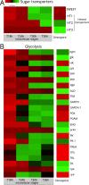
Fig. 4. Expression levels of genes involved in sugar transport and glycolysis of the marine parasite Amoebophrya across different intracellular stages within its host (the dinoflagellate Scrippsiella acuminata) and in dinospores (extracellular).
A Heatmap showing the expression level of four genes of the parasite encoding putative sugar transporters during the infection (T18 h, T24 h, T30 h and T36 h) and the dinospore stage: one SWEET (Sugars Will Eventually be Exported Transporters) and three hexose transporters (HT1, HT2, and HT3). (See also Figs. S6, S7 and S9, Table S2). B Heatmap showing the expression level of genes of the glycolysis pathway of the parasite during the infection (T18 h, T24 h, T30 h and T36 h) and in dinospores. The list of genes, their sequences and expression values can be found in Table S2.
Fig. 5
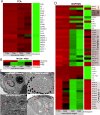
Fig. 5. Expression levels of genes involved in mitochondrial respiration and formation of cristae in the parasite Amoebophrya across different intracellular stages within its host (the dinoflagellate Scrippsiella acuminata) and in dinospores (extracellular).
Heatmap showing the expression level of genes of the TCA cycle (A) and the OXPHOS (D) pathway of the parasite. See also Fig. S10 and Table S2; (B) Expression levels of genes encoding MiC60 from the MICOS complex (MItochondrial contact site and Cristae Organizing System), and the prohibitin Phb1 and Phb2 genes. C Transmission Electron microscopy (TEM) micrographs showing the internal morphology of the mitochondrion of the parasite at different infection stages. In the cytoplasmic parasite, the electron dense mitochondrion harbored cristae (internal invagination of the inner mitochondrial membrane), which were absent in the mitochondrion of the nuclear trophont parasites (young and mature trophonts). Some vesicles could be observed in the mitochondrion of the mature trophont. Cristae reappeared in the sporont stage where the mitochondrion was substantially developed. The list of genes, their sequences and expression values can be found in Table S2.
Fig. 6
Fig. 6. Schematic overview of the potential metabolism of the marine parasite Amoebophrya inside its microalgal dinoflagellate host Scrippsiella acuminata underlying major metabolic shifts.
Major metabolic pathways have been displayed where the color of individual enzymes indicates the stage with maximum expression of their genes: yellow for intracellular parasites; blue in dinospores (free-living stage); gray whereby no difference in expression between the two stages. Dashed lines represent transport of various components; filled lines represent enzymatic reactions. Coenzymes are color-coded as purple: adenosine bi- and triphosphate (ADP and ATP, respectively); nicotinamide adenine dinucleotide phosphate (NAD(P)H); coenzyme A (CoA); quinone pool (Q and QH2). The putative location of one of the NADH:ubiquinone oxidoreductase (NDH1b/CI’) complexes, the hexose transporter (HT1) and Sugars Will Eventually be Exported Transporter (SWEET) are depicted by yellow. A question mark in the tricarboxylic acid cycle (TCA) illustrates the apparent loss of the oxoglutarate dehydrogenase/ (OGDC) or α-ketoglutarate dehydrogenase complex in Amoebophrya. Invagination of the cytoplasm to form a cytopharynx is represented to illustrate captured host chromosome to be digested in vacuoles. The list of genes, their sequences and expression values can be found in Table S2.

References

    1. Worden AZ, Follows MJ, Giovannoni SJ, Wilken S, Zimmerman AE, Keeling PJ. Rethinking the marine carbon cycle: Factoring in the multifarious lifestyles of microbes. Science. 2015;347:1257594–1257594. - PubMed
    1. Guillou L, Viprey M, Chambouvet A, Welsh RM, Kirkham AR, Massana R, et al. Widespread occurrence and genetic diversity of marine parasitoids belonging to Syndiniales (Alveolata) Environ Microbiol. 2008;10:3349–65. - PubMed
    1. Jephcott TG, Alves-de-Souza C, Gleason FH, van Ogtrop FF, Sime-Ngando T, Karpov SA, et al. Ecological impacts of parasitic chytrids, syndiniales and perkinsids on populations of marine photosynthetic dinoflagellates. Fungal Ecol. 2016;19:47–58.
    1. Alacid E, Reñé A, Garcés E. New Insights into the Parasitoid Parvilucifera sinerae Life Cycle: The Development and Kinetics of Infection of a Bloom-forming Dinoflagellate Host. Protist. 2015;166:677–99. - PubMed
    1. Not F, Gausling R, Azam F, Heidelberg JF, Worden AZ. Vertical distribution of picoeukaryotic diversity in the Sargasso Sea. Environ Microbiol. 2007;9:1233–52. - PubMed

Publication types