Review
. 2022 Oct 6:38:375-394.
doi: 10.1146/annurev-cellbio-120320-033250. Epub 2022 Jul 8.

Morphogenetic Roles of Hydrostatic Pressure in Animal Development

Affiliations
Review

Morphogenetic Roles of Hydrostatic Pressure in Animal Development

Michel Bagnat et al. Annu Rev Cell Dev Biol. .

Abstract

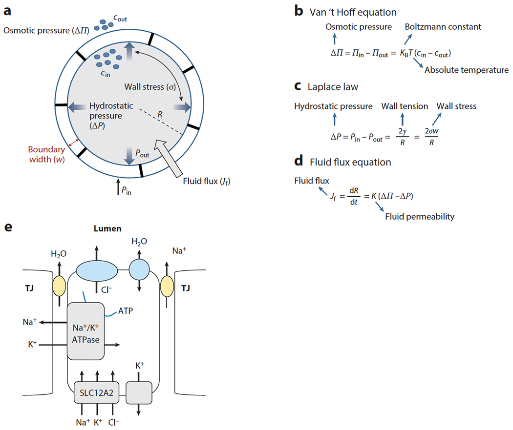
During organismal development, organs and systems are built following a genetic blueprint that produces structures capable of performing specific physiological functions. Interestingly, we have learned that the physiological activities of developing tissues also contribute to their own morphogenesis. Specifically, physiological activities such as fluid secretion and cell contractility generate hydrostatic pressure that can act as a morphogenetic force. Here, we first review the role of hydrostatic pressure in tube formation during animal development and discuss mathematical models of lumen formation. We then illustrate specific roles of the notochord as a hydrostatic scaffold in anterior-posterior axis development in chordates. Finally, we cover some examples of how fluid flows influence morphogenetic processes in other developmental contexts. Understanding how fluid forces act during development will be key for uncovering the self-organizing principles that control morphogenesis.

Keywords: axis elongation; hydrostatic pressure; lumen; morphogenesis; notochord; tube formation.


Figures

Figure 1
Figure 1
Physical and physiological principles of fluid transport in epithelial tubes. (a) Schematic of basic parameters ruling fluid flux in epithelial tubes. The symbols cin/cout and Pin/Pout are the solute concentrations and pressures inside and outside the lumen, respectively. R is the lumen radius, and w is the width of the epithelial wall. (bd) Basic parameters and laws ruling fluid flux. (e) Schematic of water and ion transport across an epithelial layer. Water may be transported via the paracellular or transcellular route. TJ stands for tight junction.
Figure 2
Figure 2
Expansion and coalescence of multiple lumina. (a) In the zebrafish gut, multiple lumina form, expand via the paracellular transport of ions and water, and coalesce into an intermediate with a double lumen or large lumina. Lumen resolution requires contact remodeling. Similar processes occur in the mammalian mammary and salivary glands (Nedvetsky et al. 2014). Panel a adapted from Alvers et al. (2014). (b) Rapid coarsening of multiple lumina and lumen expansion via transcellular transport in the zebrafish Kupffer’s vesicle (KV) and otic vesicle and in the mouse blastocyst. Epithelial thinning occurs in the zebrafish KV (A. Dasgupta et al. 2018) and otic vesicle (Hoijman et al. 2015, Mosaliganti et al. 2019). (c) Lumen expansion via mucin secretion in the Drosophila hindgut (Syed et al. 2012) and ommatidium (Husain et al. 2006).
Figure 3
Figure 3
Mathematical models of lumen formation. (a) Models of oscillatory lumen expansion behaviors. (i) Fluid flux equation. (ii) An elastic response in the mouse blastocyst produces oscillations between fixed sizes (Ruiz-Herrero et al. 2017), whereas (iii) a viscoelastic response in the zebrafish otic vesicle (Mosaliganti et al. 2019) produces oscillations with lumen size increase. (b) Flexoelectricity model. (i) Schematic of electric and surface curvature effects and mathematical expression of lumen growth. (ii) State diagram of lumen growth at different pumping and surface tension conditions showing expansion, contraction, and oscillatory behaviors. Panel b adapted from Duclut et al. (2019, 2021). (c) Schematic of the potential effect of external layers on lumen formation. If confinement (Rodriguez-Fraticelli et al. 2012) or muscle interactions (Alvers et al. 2014) reduce the stress on the epithelium, lumen formation is more efficient. Eventually it may lead to epithelial thinning (Mosaliganti et al. 2019). (d) Single lumen formation via coarsening. (i) Schematic of a multi-lumen structure. (ii) Schematic of fluid flow between microlumina of different areas (A) and tensions (γ) under isosmotic conditions (Π1 = Π2) or with a large osmotic gradient (Π1 >> Π2). (iii) Schematic of expected lumen number decay as a function of time following coarsening or Ostwald ripening. Panel d adapted from Le Verge-Serandour & Turlier (2021).
Figure 4.
Figure 4.
Hydrostatic pressure in the anterior-posterior (AP) axis of chordates. (a) Schematic of continuous lumen formation in the ascidian notochord. Lumen inflation in the notochord provides a hydrostatic scaffold for the free-swimming larva. Note that the multi-lumen intermediate resembles the organization of the vertebrate notochord. (b) Formation and inflation of fluid-filled vacuoles in the vertebrate notochord facilitates AP axis elongation. (c, top) Confocal image, (middle) 3D rendering, and (bottom) 3D arrangement of vacuolated cells in the zebrafish notochord. Vacuolated cells arrange in a stereotypical pattern (see middle panel) dictated by the relative sizes of the cells and the notochord and the aspect ratio of the tube (Norman et al. 2018). Different 3D patterns are color coded. Images in panel c provided by James Norman. (d) Inflation of vacuoles inside vacuolated cells leads to hydrostatic pressure generation inside the semirigid notochord sheath. Nuclear deformation can be used to estimate relative pressure (Bagwell et al 2020). (e) Compression of isolated vacuolated cells using atomic force microscopy and calibration of nuclear deformation may allow the determination of pressure values in vivo via noninvasive imaging. (f, top) Hydrostatic pressure in the notochord resists the compressive force of vertebral bone growth, leading to the formation of symmetrical, hourglass-shaped vertebrae. (Bottom) Loss or fragmentation of vacuoles reduces internal notochord pressure, making the structure more easily deformable and causing vertebral malformations.

References

    1. Abbas L, Whitfield TT. 2009. Nkcc1 (Slc12a2) is required for the regulation of endolymph volume in the otic vesicle and swim bladder volume in the zebrafish larva. Development 136:2837–48 - PMC - PubMed
    1. Adams DS, Keller R, Koehl MA. 1990. The mechanics of notochord elongation, straightening and stiffening in the embryo of Xenopus laevis. Development 110:115–30 - PubMed
    1. Alvers AL, Ryan S, Scherz PJ, Huisken J, Bagnat M. 2014. Single continuous lumen formation in the zebrafish gut is mediated by smoothened-dependent tissue remodeling. Development 141:1110–19 - PMC - PubMed
    1. Bagnat M, Cheung ID, Mostov KE, Stainier DY. 2007. Genetic control of single lumen formation in the zebrafish gut. Nat. Cell Biol 9:954–60 - PubMed
    1. Bagnat M, Gray RS. 2020. Development of a straight vertebrate body axis. Development 147:dev175794. - PMC - PubMed

Publication types

LinkOut - more resources