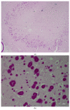
Lafora Disease and Alpha-Synucleinopathy in Two Adult Free-Ranging Moose (Alces alces) Presenting with Signs of Blindness and Circling
- PMID: 35804532
- PMCID: PMC9264765
- DOI: 10.3390/ani12131633
Lafora Disease and Alpha-Synucleinopathy in Two Adult Free-Ranging Moose (Alces alces) Presenting with Signs of Blindness and Circling
Abstract
Lafora disease is an autosomal recessive glycogen-storage disorder resulting from an accumulation of toxic polyglucosan bodies (PGBs) in the central nervous system, which causes behavioral and neurologic symptoms in humans and other animals. In this case study, brains collected from two young adult free-ranging moose (Alces alces) cows that were seemingly blind and found walking in circles were examined by light and electron microscopy. Microscopic analysis of the hippocampus of the brain revealed inclusion bodies resembling PGBs in the neuronal perikaryon, neuronal processes, and neuropil. These round inclusions measuring up to 30 microns in diameter were predominantly confined to the hippocampus region of the brain in both animals. The inclusions tested α-synuclein-negative by immunohistochemistry, α-synuclein-positive with PAS, GMS, and Bielschowsky's staining; and diastase-resistant with central basophilic cores and faintly radiating peripheral lines. Ultrastructural examination of the affected areas of the hippocampus showed non-membrane-bound aggregates of asymmetrically branching filaments that bifurcated regularly, consistent with PGBs in both animals. Additionally, α-synuclein immunopositivity was noted in the different regions of the hippocampus with accumulations of small granules ultrastructurally distinct from PGBs and morphologically compatible with alpha-synucleinopathy (Lewy body). The apparent blindness found in these moose could be related to an injury associated with secondary bacterial invasion; however, an accumulation of neurotoxicants (PGBs and α-synuclein) in retinal ganglions cells could also be the cause. This is the first report demonstrating Lafora disease with concurrent alpha-synucleinopathy (Lewy body neuropathy) in a non-domesticated animal.
Keywords: Lafora disease; Lewy body; blindness; circling; moose; polyglucosan bodies; synucleinopathies; α-Synuclein.
Conflict of interest statement
The authors declare no conflict of interest.
Figures


References
-
- Lafora G.R. Über das Vorkommen amyloider Körperchen im Innern der Ganglienzellen. Virchows Arch. Pathol. Anat. Physiol. Klin. Med. 1911;205:295–303. doi: 10.1007/BF01989438. - DOI
-
- Chan E.M., Bulman D.E., Paterson A.D., Turnbull J., Andermann E., Andermann F., Rouleau G.A., Delgado-Escueta A.V., Scherer S.W., Minassian B.A. Genetic mapping of a new Lafora progressive myoclonus epilepsy locus (EPM2B) on 6p22. J. Med. Genet. 2003;40:671–675. doi: 10.1136/jmg.40.9.671. - DOI - PMC - PubMed
-
- Ganesh S., Delgado-Escueta A.V., Suzuki T., Francheschetti S., Riggio C., Avanzini G., Rabinowicz A., Bohlega S., Bailey J., Alonso M.E., et al. Genotype-phenotype correlations for EPM2A mutations in Lafora’s progressive myoclonus epilepsy: Exon 1 mutations associate with an early-onset cognitive deficit subphenotype. Hum. Mol. Genet. 2002;11:1263–1271. doi: 10.1093/hmg/11.11.1263. - DOI - PubMed
LinkOut - more resources
Full Text Sources

