

Uterine Inflammation Changes the Expression of Cholinergic Neurotransmitters and Decreases the Population of AChE-Positive, Uterus-Innervating Neurons in the Paracervical Ganglion of Sexually Mature Gilts
- PMID: 35804576
- PMCID: PMC9264917
- DOI: 10.3390/ani12131676
Uterine Inflammation Changes the Expression of Cholinergic Neurotransmitters and Decreases the Population of AChE-Positive, Uterus-Innervating Neurons in the Paracervical Ganglion of Sexually Mature Gilts
Abstract
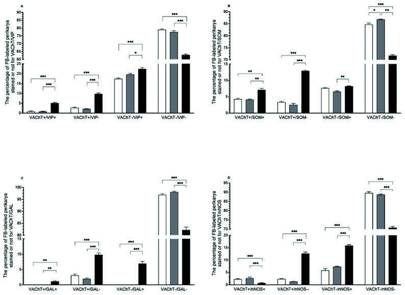
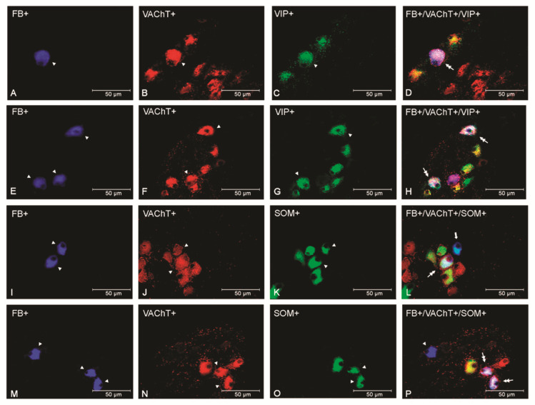
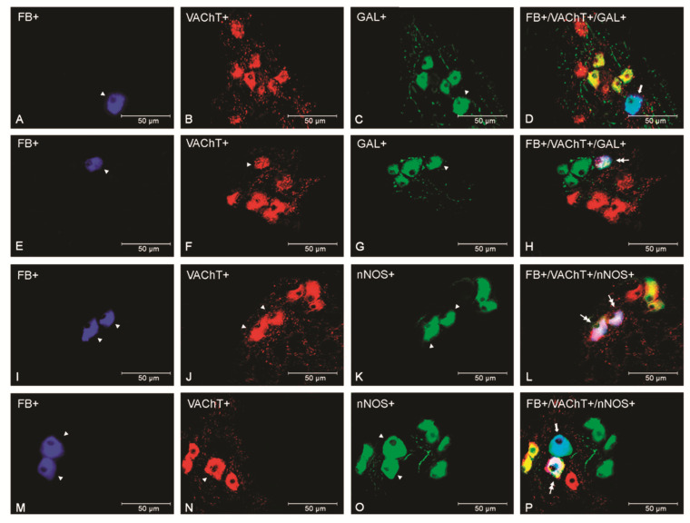
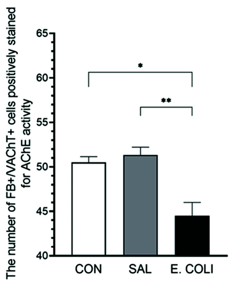
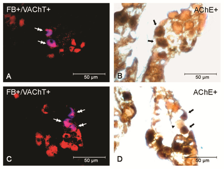
The focus of this study was based on examining the impact of endometritis on the chemical coding of the paracervical ganglion (PCG) perikaryal populations supplying pig uterus. Four weeks after the injection of Fast Blue retrograde tracer into uterine horns, either the Escherichia coli (E. coli) suspension or saline solution was applied to both horns. Laparotomy treatment was performed for the control group. Uterine cervices containing PCG were extracted on the eighth day after previous treatments. Subsequent macroscopic and histopathologic examinations acknowledged the severe form of acute endometritis in the E. coli-treated gilts, whereas double-labeling immunofluorescence procedures allowed changes to be analyzed in the PCG perikaryal populations coded with vesicular acetylcholine transporter (VAChT) and/or somatostatin (SOM), vasoactive intestinal polypeptide (VIP), a neuronal isoform of nitric oxide synthase (nNOS), galanin (GAL). The acetylcholinesterase (AChE) detection method was used to check for the presence and changes in the expression of this enzyme and further confirm the presence of cholinergic perikarya in PCG. Treatment with E. coli resulted in an increase in VAChT+/VIP+, VAChT+/VIP-, VAChT+/SOM+, VAChT+/SOM-, VAChT+/GAL- and VAChT+/nNOS- PCG uterine perikarya. An additional increase was noted in the non-cholinergic VIP-, SOM- and nNOS-immunopositive populations, as well as a decrease in the number of cholinergic nNOS-positive perikarya. Moreover, the population of cholinergic GAL-expressing perikarya that appeared in the E. coli-injected gilts and E. coli injections lowered the number of AChE-positive perikarya. The neurochemical characteristics of the cholinergic uterine perikarya of the PCG were altered and influenced by the pathological state (inflammation of the uterus). These results may indicate the additional influence of such a state on the functioning of this organ.
Keywords: chemical coding; cholinergic innervation; endometritis; immunocytochemistry; paracervical ganglion; pig; uterine neurons.
Conflict of interest statement
The authors declare no conflict of interest.
Figures
Similar articles
-
Endometritis decreases the population of uterine neurons in the paracervical ganglion and changes the expression of sympathetic neurotransmitters in sexually mature gilts.BMC Vet Res. 2021 Jul 10;17(1):240. doi: 10.1186/s12917-021-02949-z. BMC Vet Res. 2021. PMID: 34246257 Free PMC article.
-
Long-term estradiol-17β administration changes the population of paracervical ganglion neurons supplying the ovary in adult gilts.J Mol Neurosci. 2013 Jul;50(3):424-33. doi: 10.1007/s12031-012-9950-y. Epub 2013 Jan 18. J Mol Neurosci. 2013. PMID: 23329259
-
Long-term testosterone administration affects the number of paracervical ganglion ovary-projecting neurons in sexually mature gilts.Neurosci Res. 2014 Jun;83:89-96. doi: 10.1016/j.neures.2014.02.008. Epub 2014 Feb 23. Neurosci Res. 2014. PMID: 24572298
-
Endometritis Changes the Neurochemical Characteristics of the Caudal Mesenteric Ganglion Neurons Supplying the Gilt Uterus.Animals (Basel). 2020 May 20;10(5):891. doi: 10.3390/ani10050891. Animals (Basel). 2020. PMID: 32443879 Free PMC article.
-
Endometritis affects chemical coding of the dorsal root ganglia neurons supplying uterus in the sexually mature gilts.Res Vet Sci. 2019 Jun;124:417-425. doi: 10.1016/j.rvsc.2019.05.003. Epub 2019 May 7. Res Vet Sci. 2019. PMID: 31078789
References
-
- Wasowicz K., Podlasz P., Czaja K., Łakomy M. Uterus-innervating neurones of paracervical ganglion in the pig: Immunohistochemical characteristics. Folia Morphol. 2002;61:15–20. - PubMed
-
- Majewski M. Afferent and efferent innervation of the porcine ovary-sourcesof origin and chemical coding. Acta Acad. Agric. Techcol. Olst. Vet. Suppl. 1997;B24:3–125. (In Polish)
Grants and funding
LinkOut - more resources
Full Text Sources
