

Computed Tomography-Guided Fine Needle Biopsies of Vertebral and Paravertebral Lesions in Small Animals
- PMID: 35804586
- PMCID: PMC9265075
- DOI: 10.3390/ani12131688
Computed Tomography-Guided Fine Needle Biopsies of Vertebral and Paravertebral Lesions in Small Animals
Abstract
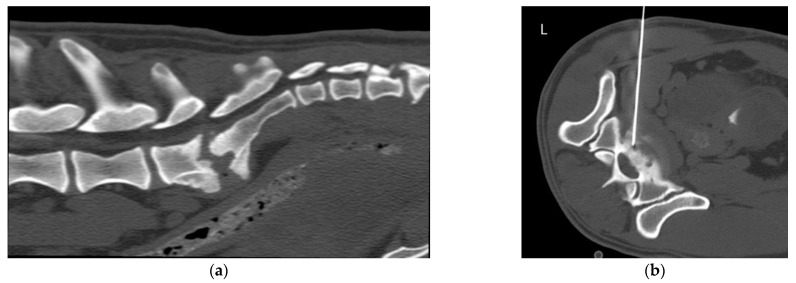
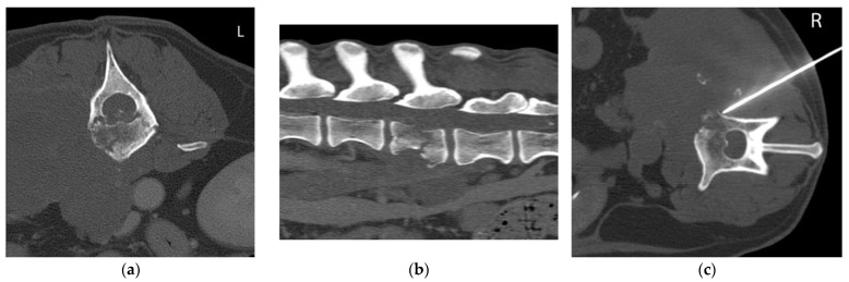
Fine needle biopsy (FNB) is an effective, minimally invasive and inexpensive diagnostic technique. Under computed tomography (CT)-guidance, lesions that have a difficult approach can be sampled to reach a diagnosis. The aim of this study is to describe the use of CT-guidance to obtain FNB from vertebral and paravertebral lesions in small animals. Ten dogs and one ferret that had undergone CT-guided FNB of vertebral and paravertebral lesions and had a cytological or a histological diagnosis were included in this retrospective study. The FNB samples were taken in four cases from the vertebra, in two cases from the intervertebral disc and in five cases from the intervertebral foramen. Two infectious and nine neoplastic lesions were diagnosed. The percentage of successful FNB was 91%. The percentage of samples with a cytological diagnosis was 80%. The percentage of complications was 9%. Limitations were the small number of animals in the study, the lacking complementary percutaneous biopsies for comparison, the lacking final histological diagnoses in some cases and the intervention of multiple operators. Computed tomography-guided FNB is a useful and safe technique for the diagnosis of vertebral and paravertebral lesions in small animals. However, a degree of expertise is important.
Keywords: computed tomographic-guidance; fine needle biopsy; small animals; vertebral.
Conflict of interest statement
The authors declare no conflict of interest.
Figures


Similar articles
-
Fine Needle Aspiration versus Fine Needle Biopsy of Biliopancreatic Lesions: Are They Really Opposing Techniques or Can They Be Complementary? Our Experience in a Large Cohort of Cases from a Single Institution.Acta Cytol. 2021;65(1):40-47. doi: 10.1159/000510755. Epub 2020 Oct 23. Acta Cytol. 2021. PMID: 33099544
-
CT-guided fine-needle biopsy of focal lung lesions as the method for reducing the number of invasive diagnostic procedures.Pol J Radiol. 2010 Apr;75(2):55-7. Pol J Radiol. 2010. PMID: 22802777 Free PMC article.
-
Comparing endoscopic ultrasound (EUS)-guided fine needle aspiration (FNA) versus fine needle biopsy (FNB) in the diagnosis of solid lesions: study protocol for a randomized controlled trial.Trials. 2016 Apr 12;17:198. doi: 10.1186/s13063-016-1316-2. Trials. 2016. PMID: 27071386 Free PMC article. Clinical Trial.
-
Endoscopic ultrasound fine needle aspiration vs fine needle biopsy for pancreatic masses, subepithelial lesions, and lymph nodes.World J Gastroenterol. 2021 Jul 14;27(26):4194-4207. doi: 10.3748/wjg.v27.i26.4194. World J Gastroenterol. 2021. PMID: 34326619 Free PMC article. Review.
-
Current role of computed tomography-guided transthoracic needle biopsy of metastatic lung lesions.Future Oncol. 2015;11(2 Suppl):43-6. doi: 10.2217/fon.14.258. Future Oncol. 2015. PMID: 25662328 Review.
Cited by
-
Clinical features, comparative imaging findings, treatment, and outcome in dogs with discospondylitis: A multi-institutional retrospective study.J Vet Intern Med. 2023 Jul-Aug;37(4):1438-1446. doi: 10.1111/jvim.16785. Epub 2023 Jun 8. J Vet Intern Med. 2023. PMID: 37288966 Free PMC article.
-
Retrospective evaluation of computed tomographic-guided Tru-Cut biopsies in 16 dogs and 14 cats with nasal cavity mass lesions.J Vet Intern Med. 2025 Jan-Feb;39(1):e17296. doi: 10.1111/jvim.17296. J Vet Intern Med. 2025. PMID: 39739338 Free PMC article.
-
Case report: MRI and CT imaging features of a melanocytic tumour affecting a cervical vertebra in an adult dog, and review of differential diagnosis for T1W-hyperintense lesions.Front Vet Sci. 2024 Apr 9;11:1334813. doi: 10.3389/fvets.2024.1334813. eCollection 2024. Front Vet Sci. 2024. PMID: 38655532 Free PMC article.
References
-
- Tidwell A.S., Johnson K.L. Computed Tomography-Guided Percutaneous Biopsy: Criteria for Accurate Needle Tip Identification. Vet. Radiol. Ultrasound. 1994;35:440–444. doi: 10.1111/j.1740-8261.1994.tb02069.x. - DOI
-
- Tidwell A.S., Johnson K.L. Computed Tomography-Guided Percutaneous Biopsy in the Dog and Cat: Description of Technique and Preliminary Evaluation in 14 Patients. Vet. Radiol. Ultrasound. 1994;35:445–456. doi: 10.1111/j.1740-8261.1994.tb02070.x. - DOI
-
- Vignoli M., Tamburro R., Felici A., del Signore F., Dettori A., di Tommaso M., Ghiraldelli A., Terragni R., Simeoni F., Falerno I., et al. Clinical Value of CT-Guided Fine Needle Aspiration and Tissue-Core Biopsy of Thoracic Masses in the Dog and Cat. Animals. 2021;11:883. doi: 10.3390/ani11030883. - DOI - PMC - PubMed
Grants and funding
LinkOut - more resources
Full Text Sources
