Review
. 2022 Jun 21;14(13):3047.
doi: 10.3390/cancers14133047.

Theranostic Potentials of Gold Nanomaterials in Hematological Malignancies

Affiliations
Review

Theranostic Potentials of Gold Nanomaterials in Hematological Malignancies

Md Salman Shakil et al. Cancers (Basel). .

Abstract

Hematological malignancies (HMs) are a heterogeneous group of blood neoplasia generally characterized by abnormal blood-cell production. Detection of HMs-specific molecular biomarkers (e.g., surface antigens, nucleic acid, and proteomic biomarkers) is crucial in determining clinical states and monitoring disease progression. Early diagnosis of HMs, followed by an effective treatment, can remarkably extend overall survival of patients. However, traditional and advanced HMs' diagnostic strategies still lack selectivity and sensitivity. More importantly, commercially available chemotherapeutic drugs are losing their efficacy due to adverse effects, and many patients develop resistance against these drugs. To overcome these limitations, the development of novel potent and reliable theranostic agents is urgently needed to diagnose and combat HMs at an early stage. Recently, gold nanomaterials (GNMs) have shown promise in the diagnosis and treatment of HMs. Magnetic resonance and the surface-plasmon-resonance properties of GNMs have made them a suitable candidate in the diagnosis of HMs via magnetic-resonance imaging and colorimetric or electrochemical sensing of cancer-specific biomarkers. Furthermore, GNMs-based photodynamic therapy, photothermal therapy, radiation therapy, and targeted drug delivery enhanced the selectivity and efficacy of anticancer drugs or drug candidates. Therefore, surface-tuned GNMs could be used as sensitive, reliable, and accurate early HMs, metastatic HMs, and MRD-detection tools, as well as selective, potent anticancer agents. However, GNMs may induce endothelial leakage to exacerbate cancer metastasis. Studies using clinical patient samples, patient-derived HMs models, or healthy-animal models could give a precise idea about their theranostic potential as well as biocompatibility. The present review will investigate the theranostic potential of vectorized GNMs in HMs and future challenges before clinical theranostic applications in HMs.

Keywords: diagnosis; gold nanomaterials; hematological malignancies; promise and challenges; treatment.


Conflict of interest statement

The authors declare no conflict of interest.

Figures

Figure 1
Figure 1
Different shapes of gold nanomaterials. The figure is reprinted from Berardis et al. (2021) [35]. This study is under Creative Commons Attribution 4.0 International License, which permits use, sharing, adaptation, distribution, and reproduction in any medium or format, as long as you give appropriate credit to the original author(s) and the source as well as provide a link to the Creative Commons license (http://creativecommons.org/licenses/by/4.0/, accessed on 26 May 2022).
Figure 2
Figure 2
Global incidence, mortality, and frequent mutations in hematological malignancies. Overall, the incidence rate was higher compared to the death toll. The data of Figure 2 (a) adapted from Sung et al. (2021) [2], and their primary data source was GLOBOCAN 2020. The gene-mutation-frequency graph was prepared based on published data on (b) leukemia (n = 3768) [39] and (c) lymphoma (n = 150) [40] patients. Nucleophosmin 1: NPM1; DNA methyltransferase 3 alpha: DNMT3A; Fms-like tyrosine kinase 3: FLT3; Neuroblastoma RAS viral oncogene homolog: NRAS; Isocitrate dehydrogenase 2: IDH2; Tet methylcytosine dioxygenase 2: TET2; Runt-related transcription factor 1: RUNX1; Tumor protein 53: TP53; Isocitrate dehydrogenase 1: IDH1; Serine and arginine-rich splicing factor 2: SRSF2; Kirsten rat sarcoma 2 viral oncogene homolog: KRAS; CREB binding protein: CREBBP; Tyrosine-protein phosphatase non-receptor type 11: PTPN11; Janus kinase 2: JAK2; Nuclear receptor binding SET domain protein 2: NSD2; Notch receptor 2: NOTCH2; Major histocompatibility complex, class I, C: HLA-C. Data source: cBioPortal.
Figure 3
Figure 3
Molecular mechanism of hematological malignancies. Oncogenic hits in a normal blood cell produce premalignant cells. Additional oncogenic hits promote the development of malignant cells. Among the malignant cells, cancer stem cells have self-renewal capacity.
Figure 4
Figure 4
Detection of chronic lymphocytic leukemia using gold nanoparticles. Dark-field images (a) and accompanying Raman spectra (b) of Giemsa and anti-CD19 Ab-conjugated AuNPs stained CLL cells. Giemsa-stained CLL cells were not visualized after anti-CD4 Ab containing AuNPs treatment in the dark-field (c), and no peak was detected by Raman spectroscopy (d). These images indicate possible results, but do not reflect any actual experiments. Chronic lymphocytic leukemia: CCL; Cluster of differentiation: CD; Antibody: Ab; Gold nanoparticles: AuPs.
Figure 5
Figure 5
Gold-nanomaterials-based leukemia detection. FA conjugated GNMs interact with the FAR overexpressed leukemia cells. (a) Interaction between FAR with FA-Pd@AuNP can be confirmed by the colorimetric method. Enzymatic activity of FA-Pd@AuNP covert H2O2 to •OH. •OH interacts with TMB to form a blue color that can be detected at the 652 nm wavelength. (b) FA-loaded AuNPs labeled with FITC binds with FAR overexpressed leukemia cells; the interaction could be confirmed by confocal microscopy, based on the green fluorescence of the FITC reporter. These images indicate potential results, but they do not reflect any actual experiments. Gold nanomaterials: GNMs; Folic acid: FA; Folic-acid receptor: FAR; Palladium gold nanoparticles: Pd@AuNPs; Gold nanoparticles: AuNPs; Fluorescein isothiocyanate: FITC.
Figure 6
Figure 6
Detection of genetic changes in leukemia using AuNPs. Interaction with the target DNA sequence with the probe DNA cause changes in Raman spectra (a) and electrochemical signal (b). Signal-enhancer molecules accelerate signal changes upon binding of target nucleic-acid moiety, thereby making changes more visible and distinguishable. These images indicate potential results, but they do not reflect any actual experiment.
Figure 7
Figure 7
Schematic representation of GNM-mediated PTT or PDT effects. Apt- or Ab-conjugated GNM binds selectively with the cancer cells. GNM or the Ptz absorb the NIR laser light. Different cellular events involved in cell death induced by GNM-mediated PTT or PDT effects upon photoexcitation. Photothermal therapy: PTT; Photodynamic therapy: PDT; Gold nanomaterials: GNM; Near-infrared radiation: NIR; Antibody: Ab; Aptamer: Apt; Reactive oxygen species: ROS.
Figure 8
Figure 8
AuNPs-based BCR-ABL gene silencing. (1) AuNPs functionalized with the e14a2 antisense hairpin ssDNA oligonucleotide (AuNP-PEG-e14a2) internalized by K562 cells, a CML in vitro model. (2) The nanoconjugate recognized BCR-ABL1 mRNA and induced silenced-gene expression and triggered mRNA degradation, thereby inhibiting (3) tyrosine kinase. The nanoconjugate (4) upregulated Bax and caspase-3, while it downregulated BCL2 expression. (5) AuNP-PEG-e14a2 increased apoptosis, resulting in decreased cell proliferation and survival. (6) IM combined with AuNP-PEG--e14a2 could be used to overcome chemoresistance. The idea of this figure was reprinted from Vinhas et al. (2017) [113]. Gold nanoparticles: AuNPs; Imatinib: IM.
Figure 9
Figure 9
Multifunctional AuNPs target miR-221 network as well as P-gp. These multifunctional AuNPs can overcome P-gp-mediated multidrug-resistance in leukemia cells. The figure is reprinted from Deng et al. (2019) [120]. This study is under Creative Commons Attribution 4.0 International License, which permits use, sharing, adaptation, distribution, and reproduction in any medium or format, as long as you give appropriate credit to the original author(s) and the source, provide a link to the Creative Commons license (http://creativecommons.org/licenses/by/4.0/, accessed on 21 December 2021). Gold nanoparticles: AuNPs; Polyethylene glycol: PEG, Folic acid: FA; Doxorubicin: Dox; Nucleolin: NCL.
Figure 10
Figure 10
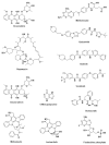
Structure of chemotherapeutic drugs used in HMs treatment. GNMs-conjugation improved the anticancer activity and selectivity of the chemotherapeutic drugs. Hematological malignancies: HMs; Gold nanomaterials: GNMs.
Figure 11
Figure 11
Response of antiangiogenic peptide-functionalized AuNPs in chorioallantoic-membrane model (CAM). Control areas are treated with PBS, K562 cells (50 μg/mL) exosome, fibroblasts (50 μg/mL) exosome, K562 exosomes (50 μg/mL) and antiangiogenic loaded AuNPs (16.4 nM). (a) CAM-region images obtained using green channel. (b) Represented image segment used to compute newly formed branches. (c) Obtained results are presented in the whisker plots. Data were obtained from six independent experiments and normalized with corresponding CAM area at 0 h incubation after PBS exposure. The 100% normalized dotted line at newly formed vessels is considered as the control sample. *1 p-value 0.0113, *2 p-value 0.0212, and *3 p-value 0.040 compared to control. This figure is adapted from Rodrigues et al. (2019) [86], non-commercial uses of this work do not require any further permission from Dove Medical Press Limited, under the license (http://creativecommons.org/licenses/by-nc/3.0/, accessed on 29 January 2022). Gold nanoparticles: AuNPs; Chorioallantoic membrane: CAM; Phosphate buffer saline: PBS.
Figure 12
Figure 12
Mechanism of GNMs recrystallization and degradation process. This summary diagram of GNMs life cycle was reprinted from Balfourier et al. (2020) [203]. The mechanism of GNMs clearance was predicated based on in vitro experimental results in human-skin primary-fibroblasts cells [203]. This study is published under PNAS license (https://www.pnas.org/authors/fees-and-licenses, accessed on 22 September 2021), permission was sincerely taken from PNAS. It is noted that stoichiometric coefficients and H+ are not presented here for clarity.

References

    1. Button E., Chan R.J., Chambers S., Butler J., Yates P. A systematic review of prognostic factors at the end of life for people with a hematological malignancy. BMC Cancer. 2017;17:213. doi: 10.1186/s12885-017-3207-7. - DOI - PMC - PubMed
    1. Sung H., Ferlay J., Siegel R.L., Laversanne M., Soerjomataram I., Jemal A., Bray F. Global cancer statistics 2020: GLOBOCAN estimates of incidence and mortality worldwide for 36 cancers in 185 countries. CA Cancer J. Clin. 2021;71:209–249. doi: 10.3322/caac.21660. - DOI - PubMed
    1. Lapotko D., Lukianova E., Shnip A., Zheltov G., Potapnev M., Savitsky V., Klimovich O., Oraevsky A. Laser activated nanothermolysis of leukemia cells monitored by photothermal microscopy; Proceedings of the Photons Plus Ultrasound: Imaging and Sensing 2005: The Sixth Conference on Biomedical Thermoacoustics, Optoacoustics, and Acousto-Optics; San Jose, CA, USA. 25 April 2005; pp. 82–89.
    1. Koutsi A., Vervesou E.C. Diagnostic molecular techniques in haematology: Recent advances. Ann. Transl. Med. 2018;6:242. doi: 10.21037/atm.2018.05.30. - DOI - PMC - PubMed
    1. Morgan G., Pratt G. Modern molecular diagnostics and the management of haematological malignancies. Clin. Lab. Haematol. 1998;20:135–141. doi: 10.1046/j.1365-2257.1998.00141.x. - DOI - PubMed

LinkOut - more resources