Review
. 2022 Jun 27;14(13):3128.
doi: 10.3390/cancers14133128.

Dynamic Interactions between Tumor Cells and Brain Microvascular Endothelial Cells in Glioblastoma

Affiliations
Review

Dynamic Interactions between Tumor Cells and Brain Microvascular Endothelial Cells in Glioblastoma

Erika Testa et al. Cancers (Basel). .

Abstract

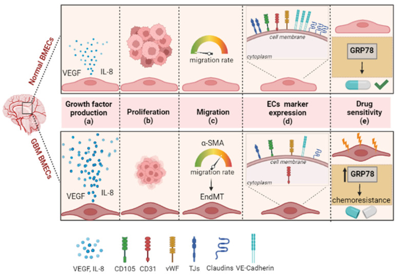
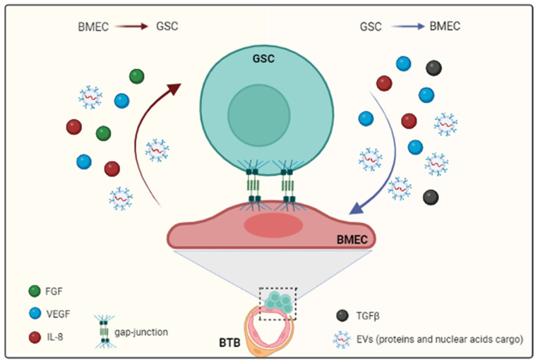
GBM is the most aggressive brain tumor among adults. It is characterized by extensive vascularization, and its further growth and recurrence depend on the formation of new blood vessels. In GBM, tumor angiogenesis is a multi-step process involving the proliferation, migration and differentiation of BMECs under the stimulation of specific signals derived from the cancer cells through a wide variety of communication routes. In this review, we discuss the dynamic interaction between BMECs and tumor cells by providing evidence of how tumor cells hijack the BMECs for the formation of new vessels. Tumor cell-BMECs interplay involves multiple routes of communication, including soluble factors, such as chemokines and cytokines, direct cell-cell contact and extracellular vesicles that participate in and fuel this cooperation. We also describe how this interaction is able to modify the BMECs structure, metabolism and physiology in a way that favors tumor growth and invasiveness. Finally, we briefly reviewed the recent advances and the potential future implications of some high-throughput 3D models to better understanding the complexity of BMECs-tumor cell interaction.

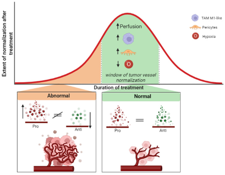
Keywords: angiogenesis; cancer; endothelial cells; extracellular vesicles; miRNA; neovascularization; tumor vessels normalization.


Conflict of interest statement

The authors declare no conflict of interest.

Figures

Figure 1
Figure 1
Schematic representation of distinct phenotypic and molecular hallmarks between normal and GBM-associated BMECs. At phenotypic level, GBM-associated BMECs show flat appearance with large nuclei, abundant cytoplasm and veil-like structures (see text for more details). In general, GBM-associated BMECs also change their intrinsic properties by increasing growth factor production such as VEGF and interleukin-8 (IL-8) (blue dots) (a); GBM-associated BMECs present a lower proliferation rate than normal BMECs (b); the migratory ability is increased in GBM-associated BMECs. The increased expression of some migration factors such as α-SMA and the interaction with brain tumoral cells lead to the endothelial-to-mesenchymal transition (EndMT) process in these cells (c); moreover, GBM-associated BMECs show the typical endothelial markers such as vWF and CD105—similarly to normal BMECs— but with a different expression level of VE-cadherin and of TJs and Claudins. More interestingly, the GBM-associated BMECs present some differences in the localization of the CD31, an endothelial cell marker, which is mainly localized into the cytoplasm rather than on the surface membrane (d); molecular alterations and the acquisition of intrinsic feature (thunders) lead to the chemoresistance in the GBM-associated BMECs. GRP78 overexpression in GBM-associated BMECs has been shown to confer chemoresistance to several drugs (two-tone pill) used in GBM treatment (e).
Figure 2
Figure 2
GSCs and BMECs communication routes. GBM induces modifications of the physical and metabolic properties of the BBB, becoming a BTB. In the resulting BTB, BMECs become the main partners of GSCs and their communication follows dynamic and bi-directional routes. This interaction occurs by direct cell contact (gap junction) or by paracrine signaling. The secreted effector molecules are growth factors (VEGF, FGF and TGF-β) and cytokines such as IL-8. In addition, EVs, in which proteins and nuclear acids are the main cargo, are an alternative route of communication (see text for more details).
Figure 3
Figure 3
The tumor vessel normalization is one of the main mechanisms of action that drives the use of antiangiogenic therapies. Due to its extensive vascularization, treatment protocols of GBM, in addition to chemotherapeutic drugs, adopt antiangiogenic compounds. However, the efficacy of antiangiogenic compounds on tumor vessels normalization seems dose- and duration-dependent. At the initial stage of the treatment, abnormal tumor vessels are a hallmark of GBM. Then, in the so-called “window of tumor vessel normalization”, through the balance between the pro- and antiangiogenic agents, the process of vessel normalization occurs. Here, the tumor vessels become normal in structure and function; the coverage of blood vessel by pericytes increases and the immune cells shift towards a tumor-associated macrophages (TAM) M1-like, leading to improved vessel perfusion and reduced tissue hypoxia. Nevertheless, the process of vessel normalization is transient and hard to capture: it occurs very quickly and lasts a short time spanning.

Similar articles

Cited by

References

    1. Obermeier B., Daneman R., Ransohoff R.M. Development, Maintenance and Disruption of the Blood-Brain Barrier. Nat. Med. 2013;19:1584–1596. doi: 10.1038/nm.3407. - DOI - PMC - PubMed
    1. Arvanitis C.D., Ferraro G.B., Jain R.K. The Blood–Brain Barrier and Blood–Tumour Barrier in Brain Tumours and Metastases. Nat. Rev. Cancer. 2020;20:26–41. doi: 10.1038/s41568-019-0205-x. - DOI - PMC - PubMed
    1. Jain R.K., di Tomaso E., Duda D.G., Loeffler J.S., Sorensen A.G., Batchelor T.T. Angiogenesis in Brain Tumours. Nat. Rev. Neurosci. 2007;8:610–622. doi: 10.1038/nrn2175. - DOI - PubMed
    1. Prager B.C., Bhargava S., Mahadev V., Hubert C.G., Rich J.N. Glioblastoma Stem Cells: Driving Resilience through Chaos. Trends Cancer. 2020;6:223–235. doi: 10.1016/j.trecan.2020.01.009. - DOI - PMC - PubMed
    1. Charles N.A., Holland E.C. The Perivascular Niche Microenvironment in Brain Tumor Progression. Cell Cycle. 2010;9:3084–3093. doi: 10.4161/cc.9.15.12710. - DOI - PMC - PubMed

LinkOut - more resources