. 2022 Jun 27;14(13):3141.
doi: 10.3390/cancers14133141.

Cancer-Associated Fibroblasts Promote Tumor Aggressiveness in Head and Neck Cancer through Chemokine Ligand 11 and C-C Motif Chemokine Receptor 3 Signaling Circuit

Affiliations

Cancer-Associated Fibroblasts Promote Tumor Aggressiveness in Head and Neck Cancer through Chemokine Ligand 11 and C-C Motif Chemokine Receptor 3 Signaling Circuit

Wen-Yen Huang et al. Cancers (Basel). .

Abstract

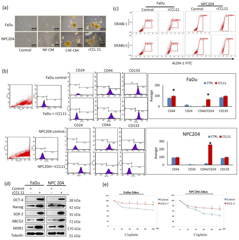
The tumor microenvironment (TME) plays a crucial role in tumor progression. One of its key stromal components, cancer-associated fibroblasts (CAFs), may crosstalk with cancer cells by secreting certain cytokines or chemokines. However, which important mediator(s) are released by CAFs, and the underlying molecular mechanism, remain largely unknown. In the present study, we isolated patient-derived CAFs and normal fibroblasts (NFs). Using microarray analysis, we detected chemokine ligand 11 (CCL11) overexpression in CAFs compared to NFs. CCL11 administration promoted the migration and invasion of head and neck cancer (HNC) cells with enhanced cancer stem cell-like properties and induction of epithelial-to-mesenchymal transition. Furthermore, neutralization of CCL11 activity reversed the aggressive phenotype of CAF-induced cancer cells. Confocal microscopy showed colocalization of CCL11 and CC chemokine receptor 3 (CCR3) on HNC cells. Moreover, immunohistochemical analysis of clinical samples from 104 patients with HNC showed that expression of CCL11 and CCR3 were significantly correlated with poor overall survival (p = 0.003 and 0.044, respectively). Collectively, CCL11 expressed on CAFs promotes HNC invasiveness, and neutralization of CCL11 reverses this effect. We propose that the CCL11/CCR3 signaling circuit is a potential target for optimizing therapeutic strategies against HNC.

Keywords: CCL11; CCR3; cancer-associated fibroblasts; head and neck cancer; tumor microenvironment.


Conflict of interest statement

The authors declare no conflict of interest.

Figures

Figure 1
Figure 1
Characterization of CAFs and NFs obtained from clinical surgical tissues from patients with head and neck cancer (HNC) (a) Morphological comparisons between CAFs and NFs from a representative HNC case showed that CAFs (right panel) consisted of more cytoplasmic protrusions than NFs (left panel). Photographs were captured at 40× magnification. (b) Quantitative PCR (left panel) of the culture medium showed a significantly higher expression of vimentin and α-SMA in CAFs than in NFs. Western blot analysis (right panel) also demonstrated that levels of vimentin and α-SMA were significantly higher in CAFs than in NFs. (c) Flow cytometric analysis of the cell surface markers, CD10 and GPR77, showed a marked increase in their expression in CAFs compared to NFs. (d) A heat map of the gene microarray of NFs and CAFs showed that there were several differences in the expression profile of the secreted genes. The arrow indicates a marked discrepancy of the relative mRNA levels of CCL11 in CAF compared with NF. (e) The RT-PCR (left panel) and ELISA (right panel) analysis showed an increased expression of CCL11 in CAFs compared with that in NFs. (f) Western blot analysis showed that the protein level of CCL11 was higher in CAFs than in NFs in cell lysates. (g) Western blot analysis showed a higher CCL11 expression in CAF-CM compared to NF-CM. The asterisk indicated a significant difference (*: p < 0.05; **: p < 0.01) between experimental and control groups.
Figure 2
Figure 2
CCL11 produced by CAFs causes increased migration and invasion, and the EMT of HNC cells. (a) Comparative analysis of the migration and invasion of HNC cells associated with CCL11. Four test groups were classified for comparative analysis of migration and invasive abilities. FaDu and NPC204 cells cultured with medium containing CAF-induced CCL11 presented greater abilities of migration and invasion, with a statistically significant difference, than three other groups: NF, NF with CCL11, and CAFs treated with CCL11 antibody. The asterisk indicates a significant difference (*: p < 0.05; **: p < 0.01) (b) Comparative photographs of the infiltrating behavior of FaDu and NPC204 cells in an organotypic culture in four groups seeded onto a mixture layer containing NFs or CAFs with CCL11 or CCL11 antibody. The arrow(s) indicate infiltration buds from the HNC cells seeded above. (c) Representative blots of the EMT-associated markers in FaDu and NPC204 cells, as observed upon Western blotting analysis in five groups, showed that treatment with CAF-conditioned medium or the application of rCCL11 decreased the expression of epithelial-type markers (E-cadherin), and increased the expression of mesenchymal-type markers (fibronectin) and EMT regulators (Snail and Twist). In addition, increased expression of invasion-related MMP2 and MMP9 was also seen in those two groups, compared with other groups. The asterisk indicates a significant difference (p < 0.05) between experimental and control groups. Results are expressed as mean ± SD.
Figure 3
Figure 3
Comparative analysis of induction of CSC properties and drug resistance in HNC cells associated with CCL11. (a) Four groups were classified for comparative analysis of the ability of sphere formation. Increased ability of sphere formation in two test groups of HNC cells exposed to a CAF medium and the group with treatment of rCCL11 was noted. (b) Flow cytometric analysis showed a significant increase in CD44 and CD44/CD24, as well as in CD133 in HNC cells exposed to rCCL11, compared to control HNC cells (p < 0.05). (c) Flow cytometric analysis showed a marked increase in ALDH-1 activity in HNC cells exposed to rCCL11 compared to control HNC cells. (d) Western blot analysis showed that CSC-representative markers, Oct-4, Nanog, and Sox-2, were also overexpressed in addition to the increased expression of two important drug resistance genes, ABCG-2 and MDR-1, in HNC cells exposed to rCCL11. (e) Treatment with Cisplatin at 24 h showed a significant increase in chemoresistance in both FaDu and NPC204 cells exposed to rCCL11 compared with control HNC cells. The asterisk indicates a significant difference (*: p < 0.05; **: p < 0.01) between experimental and control groups.
Figure 4
Figure 4
CCL11 and CCR3 expression with associated signal pathway in HNC cell lines and their correlation to clinical outcomes in 104 HNC patients. (a) Confocal microscopic images showed CCL11 (green) localized to both cell and nuclear membranes, while CCR3 (red) localized only to the cell membrane in FaDu cells; CCL11 and CCR3 co-localized at the cell membrane (yellow). In NPC204 cells, CCL11 (green) and CCR3 (red) were found to co-localize at protrusions polarized to the cells (yellow). (b) Using the crisp technique, higher expression of CCR3, MMP2, and MMP3 was found in over-expressed CCL11 cloned-FaDu and NPC204 cells. Cloned CCL11-overexpressed cells were abolished by adding eotaxin siRNA or CCR3 antibody, which reversed the expression of CCR3 and invasion-related MMP2 and MMP9. (c) Higher phosphorylation levels of p38 MAPK and ERK were found in cloned CCL11-overexpressed FaDu and NPC204 cells and were reversed by treatment of the p38 MAPK inhibitor (SB203580) and ERK inhibitor (FR180204), respectively. The phosphorylation level of JNK was kept in low condition before and after treatment of the JNK inhibitor (SP600125). (d) Photomicrographs of immunohistochemical staining from tissue microarray showing CCL11 and CCR3 expression in three different representative groups of HNC patients (magnification, ×200). (e) Kaplan–Meier survival analysis of patients showed that overexpression of CCL11 and CCR3 were statistically associated with poor overall survival).
Figure 5
Figure 5
The diagrammatic illustration demonstrates the major mechanism that CAFs secreting CCL11 promotes HNC cell migration and invasion and induces properties of drug resistance and stemness, shown as follows. CAFs in TME secret CCL11 binding to the CCR3 receptors on HNC cells via the paracrine effect. The signal induces overexpression of transcriptional factors, such as Snail and Twist, which regulate EMT and are also responsible for self-induction of CCL11 in an autocrine fashion. As a result, CCL11, via paracrine or autocrine signaling when targeting CCR3 receptors, play a functional role in the induction of EMT and CSC properties, for further tumor progression.

Similar articles

Cited by

References

    1. Bray F., Ferlay J., Soerjomataram I., Siegel R.L., Torre L.A., Jemal A. Global cancer statistics 2018: GLOBOCAN estimates of incidence and mortality worldwide for 36 cancers in 185 countries. CA Cancer J. Clin. 2018;686:394–424. doi: 10.3322/caac.21492. - DOI - PubMed
    1. Chow L.Q.M. Head and Neck Cancer. N. Engl. J. Med. 2020;3821:60–72. doi: 10.1056/NEJMra1715715. - DOI - PubMed
    1. Hamidi H., Ivaska J. Every step of the way: Integrins in cancer progression and metastasis. Nat. Rev. Cancer. 2018;189:533–548. doi: 10.1038/s41568-018-0038-z. - DOI - PMC - PubMed
    1. Peltanova B., Raudenska M., Masarik M. Effect of tumor microenvironment on pathogenesis of the head and neck squamous cell carcinoma: A systematic review. Mol. Cancer. 2019;181:63. doi: 10.1186/s12943-019-0983-5. - DOI - PMC - PubMed
    1. Gascard P., Tlsty T.D. Carcinoma-associated fibroblasts: Orchestrating the composition of malignancy. Genes Dev. 2016;309:1002–1019. doi: 10.1101/gad.279737.116. - DOI - PMC - PubMed

LinkOut - more resources