Review
. 2022 Jun 27;14(13):3147.
doi: 10.3390/cancers14133147.

Imaging of Uveal Melanoma-Current Standard and Methods in Development

Affiliations
Review

Imaging of Uveal Melanoma-Current Standard and Methods in Development

Małgorzata Solnik et al. Cancers (Basel). .

Abstract

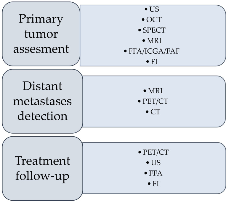
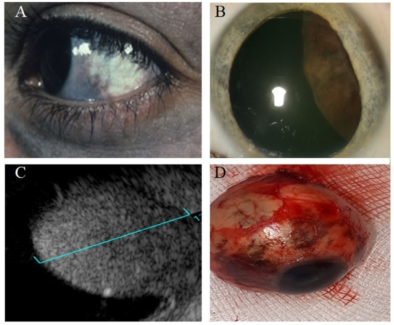
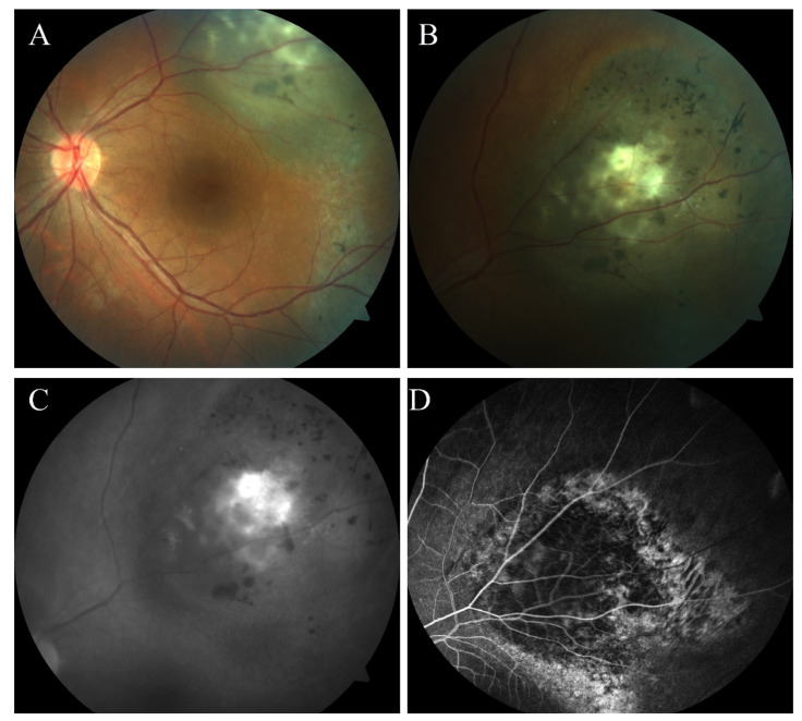
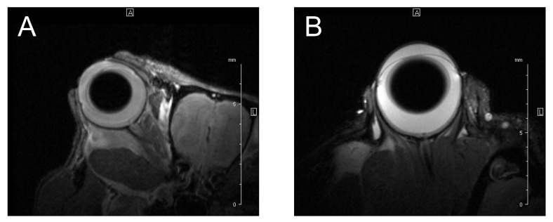
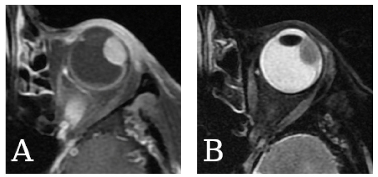
Uveal melanoma is the most common primary intraocular malignancy in adults, characterized by an insidious onset and poor prognosis strongly associated with tumor size and the presence of distant metastases, most commonly in the liver. Contrary to most tumor identification, a biopsy followed by a pathological exam is used only in certain cases. Therefore, an early and noninvasive diagnosis is essential to enhance patients' chances for early treatment. We reviewed imaging modalities currently used in the diagnostics of uveal melanoma, including fundus imaging, ultrasonography (US), optical coherence tomography (OCT), single-photon emission computed tomography (SPECT), fundus fluorescein angiography (FFA), indocyanine green angiography (ICGA), fundus autofluorescence (FAF), as well as positron emission tomography/computed tomography (PET/CT) or magnetic resonance imaging (MRI). The principle of imaging techniques is briefly explained, along with their role in the diagnostic process and a summary of their advantages and limitations. Further, the experimental data and the advancements in imaging modalities are explained. We describe UM imaging innovations, show their current usage and development, and explain the possibilities of utilizing such modalities to diagnose uveal melanoma in the future.

Keywords: CT; MRI; OCT; PET; SPECT; diagnosis; imaging; ultrasonography; uveal melanoma.


Conflict of interest statement

The authors declare no conflict of interest.

Figures

Figure 1
Figure 1
Preferable imaging techniques in different stages of diagnosis and follow-up of uveal melanoma. US—ultrasonography; OCT—optical coherence tomography; SPECT—single-photon emission computed tomography; MRI—magnetic resonance imaging; FFA—fundus fluorescein angiography; ICGA—indocyanine green angiography; FAF—fundus autofluorescence; PET—positron emission tomography; CT—computed tomography.
Figure 2
Figure 2
Choroidal melanoma: fundus photography (A), red-free image (B), OCT scan (C), and US (D).
Figure 3
Figure 3
Echographic features of choroidal melanoma. Melanotic choroidal melanoma is mushroom-shaped as detected at the level of inferior arcade of the left eye (A). That was shown to be mushroom-shaped with acoustic hollowness in B-scan (B). One-dimensional A-scan imaging (8 MHz) for an eye with choroidal melanoma (C). The vertical deflections represent echoes from different surfaces in the eye. The red arrow indicates the surface of the retina, the yellow arrow indicates the surface of the sclera, and the interval between them (blue line) shows the low reflectivity of the tumor (choroidal melanoma). Ciliary body melanoma (D) detected in ultrasound biomicroscopy (UBM).
Figure 4
Figure 4
Left eye with amelanotic choroidal melanoma (A,B) showed a 7.7 mm thick dome shape tumor with moderate low internal reflectivity. Six months after treatment with I-125 radioactive plaque, the tumor showed regression clinically (C), and thickness decreased to 4.2 mm (D).
Figure 5
Figure 5
Patient with oculo-dermal melanosis (A) was found to have retrolenticular melanotic mass (B), which was found to be large choroidal melanoma (13 mm thick) with ciliary body involvement (C). This eye was enucleated and shown to have scleral melanosis (D).
Figure 6
Figure 6
A 30-year-old female with left melanotic choroidal melanoma (A). The clinical exam showed RPE hyperplasia and metaplasia, indicating underlying previous choroidal nevus (B). The lesion has orange pigment lipofuscins that showed fundus autofluorescence (C), and the mass was associated with intrinsic vascularity in FFA (D).
Figure 7
Figure 7
T2-weighted MR images of the eyes of experimental animals—(A) mouse and (B) rat. High-resolution images enable detailed evaluation of the ocular anatomy.
Figure 8
Figure 8
T1- (A) and T2-weighted (B) MR images of the human eyes with uveal melanoma.

Similar articles

Cited by

References

    1. Kaliki S., Shields C.L. Uveal melanoma: Relatively rare but deadly cancer. Eye. 2016;31:241–257. doi: 10.1038/eye.2016.275. - DOI - PMC - PubMed
    1. Spagnolo F., Caltabiano G., Queirolo P. Uveal melanoma. Cancer Treat. Rev. 2012;38:549–553. doi: 10.1016/j.ctrv.2012.01.002. - DOI - PubMed
    1. Souto E.B., Zielinska A., Luis M., Carbone C., Martins-Gomes C., Souto S.B., Silva A.M. Uveal melanoma: Physiopathology and new in situ-specific therapies. Cancer Chemother. Pharmacol. 2019;84:15–32. doi: 10.1007/s00280-019-03860-z. - DOI - PubMed
    1. Dogrusöz M., Jager M.J., Damato B. Uveal Melanoma Treatment and Prognostication. Asia-Pac. J. Ophthalmol. 2017;6:186–196. doi: 10.22608/apo.201734. - DOI - PubMed
    1. Blum E.S., Yang J., Komatsubara K.M., Carvajal R.D. Clinical Management of Uveal and Conjunctival Melanoma. Oncology. 2016;30:29. - PubMed

LinkOut - more resources