

Preferential Antibody and Drug Conjugate Targeting of the ADAM10 Metalloprotease in Tumours
- PMID: 35804938
- PMCID: PMC9264901
- DOI: 10.3390/cancers14133171
Preferential Antibody and Drug Conjugate Targeting of the ADAM10 Metalloprotease in Tumours
Abstract
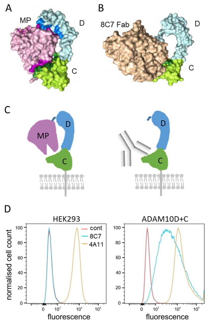
ADAM10 is a transmembrane metalloprotease that sheds a variety of cell surface proteins, including receptors and ligands that regulate a range of developmental processes which re-emerge during tumour development. While ADAM10 is ubiquitously expressed, its activity is normally tightly regulated, but becomes deregulated in tumours. We previously reported the generation of a monoclonal antibody, 8C7, which preferentially recognises an active form of ADAM10 in human and mouse tumours. We now report our investigation of the mechanism of this specificity, and the preferential targeting of 8C7 to human tumour cell xenografts in mice. We also report the development of novel 8C7 antibody-drug conjugates that preferentially kill cells displaying the 8C7 epitope, and that can inhibit tumour growth in mice. This study provides the first demonstration that antibody-drug conjugates targeting an active conformer of ADAM10, a widely expressed transmembrane metalloprotease, enable tumour-selective targeting and inhibition.
Keywords: ADAM metalloprotease; antibody–drug conjugate; brain cancer; colon cancer; therapeutic antibody.
Conflict of interest statement
P.W.J., A.M.S., N.S. and D.B.N. are inventors of granted patents relevant to the anti-ADAM10 antibody 8C7. The funders had no role in the design of the study; in the collection, analyses, or interpretation of data; in the writing of the manuscript, or in the decision to publish the results.
Figures


Similar articles
-
An activated form of ADAM10 is tumor selective and regulates cancer stem-like cells and tumor growth.J Exp Med. 2016 Aug 22;213(9):1741-57. doi: 10.1084/jem.20151095. Epub 2016 Aug 8. J Exp Med. 2016. PMID: 27503072 Free PMC article.
-
Antibodies binding the ADAM10 substrate recognition domain inhibit Eph function.J Cell Sci. 2012 Dec 15;125(Pt 24):6084-93. doi: 10.1242/jcs.112631. Epub 2012 Oct 29. J Cell Sci. 2012. PMID: 23108669 Free PMC article.
-
The TspanC8 subgroup of tetraspanins interacts with A disintegrin and metalloprotease 10 (ADAM10) and regulates its maturation and cell surface expression.J Biol Chem. 2012 Nov 16;287(47):39753-65. doi: 10.1074/jbc.M112.416503. Epub 2012 Oct 3. J Biol Chem. 2012. PMID: 23035126 Free PMC article.
-
Scissor sisters: regulation of ADAM10 by the TspanC8 tetraspanins.Biochem Soc Trans. 2017 Jun 15;45(3):719-730. doi: 10.1042/BST20160290. Biochem Soc Trans. 2017. PMID: 28620033 Free PMC article. Review.
-
Alpha-Secretase ADAM10 Regulation: Insights into Alzheimer's Disease Treatment.Pharmaceuticals (Basel). 2018 Jan 29;11(1):12. doi: 10.3390/ph11010012. Pharmaceuticals (Basel). 2018. PMID: 29382156 Free PMC article. Review.
Cited by
-
ADAM Proteases in Cancer: Biological Roles, Therapeutic Challenges, and Emerging Opportunities.Cancers (Basel). 2025 May 19;17(10):1703. doi: 10.3390/cancers17101703. Cancers (Basel). 2025. PMID: 40427200 Free PMC article. Review.
-
Abnormal expressions of PURPL, miR-363-3p and ADAM10 predicted poor prognosis for patients with ovarian serous cystadenocarcinoma.J Cancer. 2023 Sep 11;14(15):2908-2918. doi: 10.7150/jca.87405. eCollection 2023. J Cancer. 2023. PMID: 37781085 Free PMC article.
References
-
- Hartmann D., de Strooper B., Serneels L., Craessaerts K., Herreman A., Annaert W., Umans L., Lubke T., Lena Illert A., von Figura K., et al. The disintegrin/metalloprotease ADAM 10 is essential for Notch signalling but not for alpha-secretase activity in fibroblasts. Hum. Mol. Genet. 2002;11:2615–2624. doi: 10.1093/hmg/11.21.2615. - DOI - PubMed
Grants and funding
LinkOut - more resources
Full Text Sources
