Review
. 2022 Jun 29;14(13):3180.
doi: 10.3390/cancers14133180.

Imaging of Pediatric Testicular and Para-Testicular Tumors: A Pictural Review

Affiliations
Review

Imaging of Pediatric Testicular and Para-Testicular Tumors: A Pictural Review

Anne-Laure Hermann et al. Cancers (Basel). .

Abstract

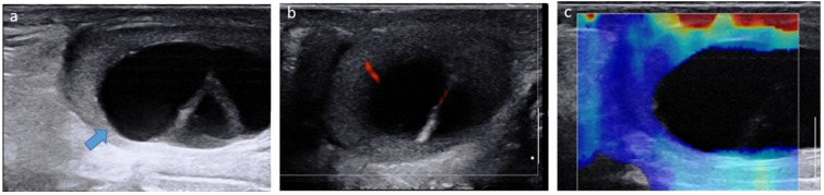
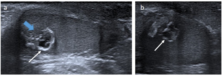
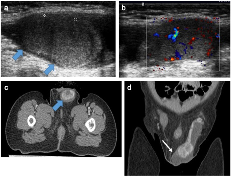
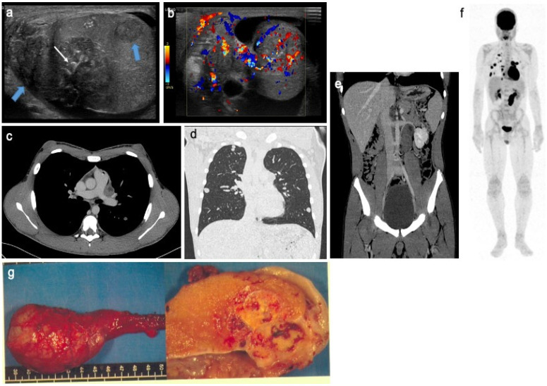
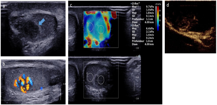
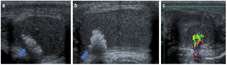
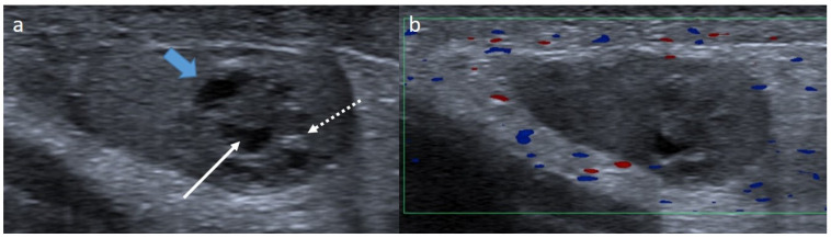
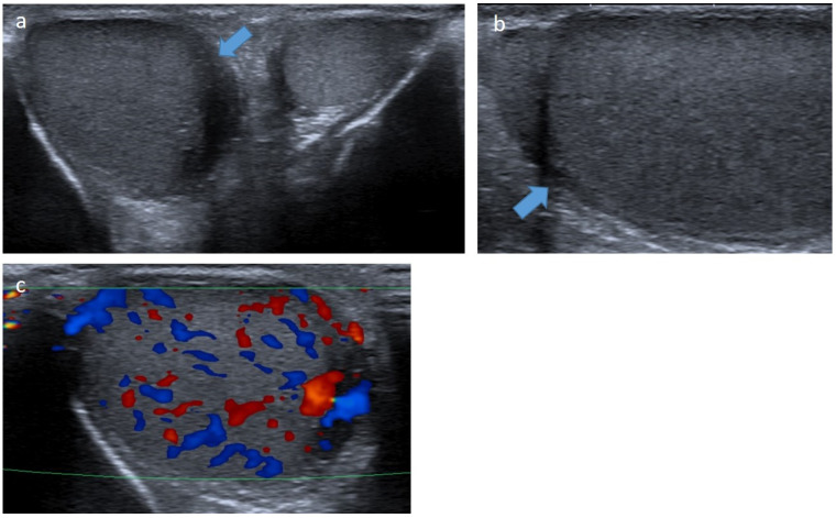
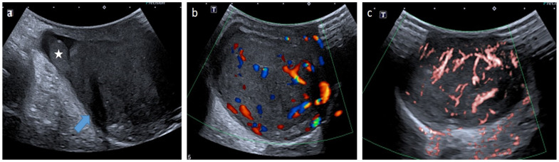
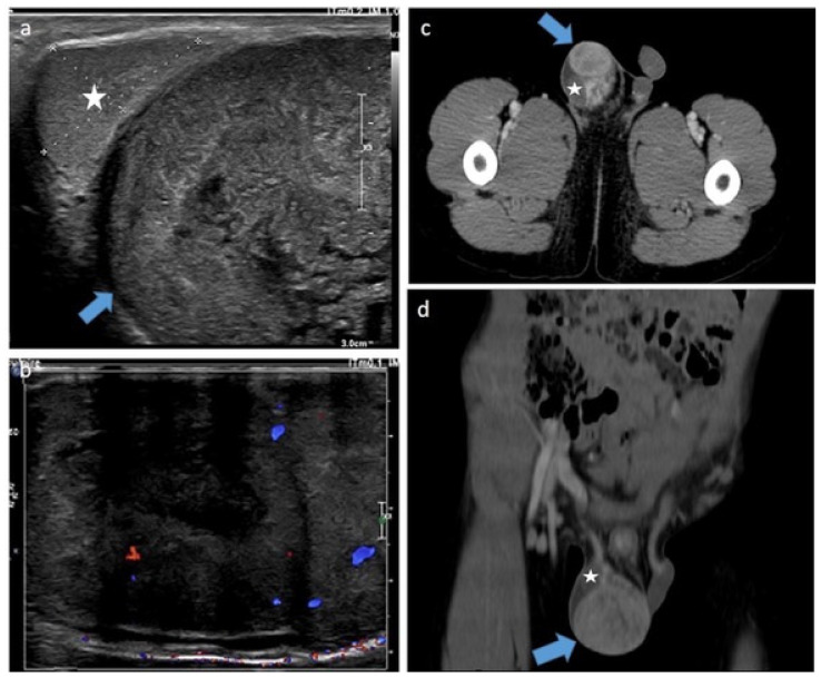
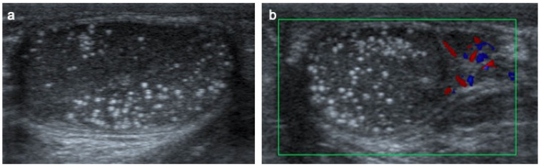
Pre- and post-pubertal testicular tumors are two distinct entities in terms of epidemiology, diagnosis and treatment. Most pre-pubertal tumors are benign; the most frequent are teratomas, and the most common malignant tumors are yolk-sac tumors. Post-pubertal tumors are similar to those found in adults and are more likely to be malignant. Imaging plays a pivotal role in the diagnosis, staging and follow-up. The appearance on ultrasonography (US) is especially helpful to differentiate benign lesions that could be candidates for testis-sparing surgery from malignant ones that require radical orchidectomy. Some specific imaging patterns are described for benign lesions: epidermoid cysts, mature cystic teratomas and Leydig-cell tumors. Benign tumors tend to be well-circumscribed, with decreased Doppler flow on US, but malignancy should be suspected when US shows an inhomogeneous, not-well-described lesion with internal blood flow. Imaging features should always be interpreted in combination with clinical and biological data including serum levels of tumor markers and even intra-operative frozen sections in case of conservative surgery to raise any concerns of malignity. This review provides an overview of imaging features of the most frequent testicular and para-testicular tumor types in children and the value of imaging in disease staging and monitoring children with testicular tumors or risk factors for testicular tumors.

Keywords: color Doppler; germ cell tumors; imaging; non-germ cell tumors; pediatric testicular tumors; pre-pubertal tumors; scrotal MRI; ultrasound.


Conflict of interest statement

The authors declare no conflict of interest.

Figures

Figure 1
Figure 1
Mature cystic teratoma in a 4-month-old boy. Longitudinal gray-scale B mode (a), color Doppler (b) and strain elastography (c) US of the testis. Images show a cystic intra-testicular lesion (blue arrow) containing two inner septa with color Doppler signals and no signals on elastography. Serum levels of tumor markers were normal. The patient underwent conservative surgery. US, ultrasonography.
Figure 2
Figure 2
Epidermoid cyst in a 20-month-old boy. Longitudinal (a) and transverse (b) gray-scale B mode US of the testis. Images show a well-demarcated intra-testicular lesion (blue arrow) with a small central anechoic part and a peripheral hyperechoic rim, giving an “onion-ring appearance” (white arrows). This lesion was incidentally discovered on scrotal US for a contralateral undescended testis. Serum levels of tumor markers were normal. The patient underwent conservative surgery. US, ultrasonography.
Figure 3
Figure 3
Intra-testicular yolk-sac tumor in a 20-month-old boy. Longitudinal gray-scale B mode (a) and transverse color Doppler (b) US of the testis. Staging contrast-enhanced CT focused on the pelvic on axial (c) and coronal (d) views. Images demonstrating two solid intra-testicular masses with color Doppler signal and heterogeneous enhancement after contrast (blue arrow), surrounded by peritesticular effusion (white arrow). AFP level was high and inguinal orchidectomy was performed. US, ultrasonography; AFP, alpha-fetoprotein. Courtesy of Dr Brisse and Dr Cardoen, Institut Curie, Paris, France.
Figure 4
Figure 4
Mixed non-seminomatous malignant germ cell tumor in a 14-year-old boy with yolk-sac and choriocarcinoma components. Longitudinal gray-scale B mode (a) and transverse color Doppler (b) US of the testis. Contrast-enhanced CT on axial view of the mediastinum (c), coronal view of the lung (d) and coronal view of the abdomen and pelvis (e). Coronal view of 18F-FDG PET (f). Images show several heterogeneous testicular lesions (blue arrow) containing micro calcifications (white arrow) and highly vascularized tissular portions with irregular distribution (b). CT scan demonstrates metastatic mediastinal and retroperitoneal lymph nodes and multiple lung metastases with high uptake on 18F-FDG PET. (g) Macroscopic view with gross pathology section of the lesion after radical surgery. This adolescent presented a rapid increase in testicular volume and increased AFP and BHCG levels. US, ultrasonography; AFP, alpha-fetoprotein; BHCG, beta-human chorionic gonadotrophin; 18F-FDG PET, 18F fluorodeoxyglucose positron emission tomography. Courtesy of Dr Brisse and Dr Cardoen, Institut Curie, Paris, France and Pr Berrebi, Robert Debré Hospital, Paris, France.
Figure 5
Figure 5
Leydig-cell tumor in an 8-year-old boy. Transverse gray-scale B-mode (a) and longitudinal color Doppler (b) US of the testis. Shear wave elastography cartography (c) and contrast-enhanced US focused on the lesion (d). Images show a well-circumscribed hypoechoic mass with intrinsic central and peripheral hypervascularization, surrounded by a hyperechoic rim (blue arrow) with moderate increased stiffness on elastography. The patient was referred for early pubic hairiness and accelerated growth rate. Testosterone level was increased, and gonadotrophin levels (follicle-stimulating hormone, luteinizing hormone) were low. The patient underwent conservative surgery. US, ultrasonography. Courtesy of Pr Laurence Rocher, Antoine Béclère, Clamart, France.
Figure 6
Figure 6
Sertoli-cell tumor in an 8-years-old boy with Peutz–Jeghers syndrome. Transverse (a), longitudinal (b) gray-scale B-mode and transverse color Doppler (c) US of the testis. Images show a hypoechoic nodule mostly calcified with irregular calcifications (blue arrow) causing posterior shadowing and artifacts on color Doppler imaging. US, ultrasonography. The child presented with acceleration of his growth and gynecomastia. Later, he developed nasal polyps and mucocutaneous pigmentation and Peutz–Jeghers syndrome was confirmed. The patient underwent conservative surgery.
Figure 7
Figure 7
Granulosa-cell tumor in a 1-month-old boy. Longitudinal gray-scale B mode (a) and color Doppler (b) US of the testis. Images show a well-circumscribed mass with lobulated margins (blue arrow) containing a cystic component (arrow) and calcifications (dotted arrow). Serum levels of tumor markers were normal, and partial orchidectomy with an inguinal approach was performed. US, ultrasonography.
Figure 8
Figure 8
Leukemic testicular infiltration of the right testis in a 10-year-old boy. Transverse (a) and longitudinal (b) gray-scale B-mode and transverse color Doppler (c) US of the testis. Images show an enlargement of the right testis demonstrating an ill-defined hypoechoic infiltration (blue arrow) with increased intralesional flow preserving the normal vascular architecture. This infiltration regressed on chemotherapy. US, ultrasonography.
Figure 9
Figure 9
Metastatic neuroblastoma with liver, bone and testis involvement. Contrast-enhanced abdominal CT scan (a), different views of gray-scale B-mode US focused on the abdomen (b), testis (c) and liver (d). MIBG scan (e). Images show a left adrenal tissular mass (white arrow) with a mass effect on the left kidney (star) and intra-testicular (blue arrow) and liver metastases. The testicular metastasis appears as a well-circumscribed hypoechoic and heterogeneous mass. In addition, MIBG scan demonstrates bone extension. US, ultrasonography; MIBG, meta-iodobenzylguanidine. Courtesy of Dr Brisse, Institut Curie, Paris, France.
Figure 10
Figure 10
Testicular adrenal rest tumors in a 6-month boy with congenital adrenal hyperplasia. Transverse gray-scale B-mode (a) and color Doppler (b) US of the testis. Images show bilateral and symmetric lesions at the hilum of the testis, mostly hypoechoic with a hyperechoic center, lobulated margins (blue arrow) and increased intralesional flow preserving the normal vascular architecture. US, ultrasonography. Courtesy of Pr Laurence Rocher, Antoine Béclère, Clamart, France.
Figure 11
Figure 11
Para-testicular dermoid cyst in a 6-year-old boy. Longitudinal (a) gray-scale B-mode and transverse color Doppler (b) US of the scrotum. Images show an extra-testicular lesion (blue arrow) at the lower pole of the testis (star), demonstrating well-circumscribed margins, homogeneous echoic content with acoustic posterior reinforcement (arrow) without internal vessels on color Doppler images. Serum levels of tumor markers were normal, and the lesion was removed by an inguinal approach. US, ultrasonography.
Figure 12
Figure 12
Para-testicular epithelial cyst in a 15-year-old boy. Longitudinal gray-scale B-mode (a) and color Doppler (b) US of the testis. Images show a small para-testicular cyst (blue arrow) in contact with albuginea. The lesion was removed with a scrotal approach, and pathology revealed a para-testicular epithelial cyst. US, ultrasonography.
Figure 13
Figure 13
Para-testicular rhabdomyosarcoma in an 8-year-old boy (case 1). Transverse gray-scale B-mode (a), color Doppler (b) and power Doppler (c) US of the scrotum. Images show a solid lesion (blue arrow) in an extra-testicular location (testis is represented by a star), with heterogeneous aspect and showing high and irregular vascularization. Inguinal radical orchidectomy was performed, followed by a course of chemotherapy. The margins were safe. US, ultrasonography.
Figure 14
Figure 14
Para-testicular rhabdomyosarcoma in a 6-year-old boy (case 2). Transverse gray-scale B-mode (a) and color Doppler (b) US of the scrotum. Staging contrast-enhanced CT scan focused on the pelvic on axial (c) and coronal (d) views. Images show a solid heterogeneous lesion (blue lesion) in an extra-testicular location (testis is represented by a star) with internal vessels on color Doppler images and showing high and heterogeneous enhancement after contrast administration. US, ultrasonography.
Figure 15
Figure 15
Testicular microlithiasis in a 6-year-old boy. Transverse gray-scale B-mode (a) and color Doppler (b) US of the scrotum. Images show diffuse hyperechoic punctate calcifications.

References

    1. Ross J.H. Prepubertal Testicular Tumors. Urology. 2009;74:94–99. doi: 10.1016/j.urology.2008.12.036. - DOI - PubMed
    1. Moreno C.C., Small W.C., Camacho J.C., Master V., Kokabi N., Lewis M., Hartman M., Mittal P.K. Testicular Tumors: What Radiologists Need to Know—Differential Diagnosis, Staging, and Management. RadioGraphics. 2015;35:400–415. doi: 10.1148/rg.352140097. - DOI - PubMed
    1. Stein R., Quaedackers J., Bhat N.R., Dogan H.S., Nijman R.J., Rawashdeh Y.F., Silay M.S., Hoen L.A., Tekgul S., Radmayr C., et al. EAU-ESPU pediatric urology guidelines on testicular tumors in prepubertal boys. J. Pediatr. Urol. 2021;17:529–533. doi: 10.1016/j.jpurol.2021.06.006. - DOI - PubMed
    1. Ye Y.-L., He Q.-M., Zheng F.-F., Guo S.-J., Zhou F.-J., Qin Z.-K. Trends of testis-sparing surgery for pediatric testicular tumors in South China. BMC Surg. 2017;17:31. doi: 10.1186/s12893-017-0230-1. - DOI - PMC - PubMed
    1. Jiménez Isabel M.A., Gómez Fraile A., Aransay Brantot A., López Vázquez F., Delgado Muños M.D., Encinas Goenechea A., Matute de Cárdenas M.D., Berchi García F.J. Testicular tumors in childhood. Review of cases in the course of 13 years. Cir. Pediatr. 1996;9:13–16. - PubMed

LinkOut - more resources