. 2022 Jun 21;11(13):1984.
doi: 10.3390/cells11131984.

Standardization of Cell Culture Conditions and Routine Genomic Screening under a Quality Management System Leads to Reduced Genomic Instability in hPSCs

Affiliations

Standardization of Cell Culture Conditions and Routine Genomic Screening under a Quality Management System Leads to Reduced Genomic Instability in hPSCs

Francisco J Molina-Ruiz et al. Cells. .

Abstract

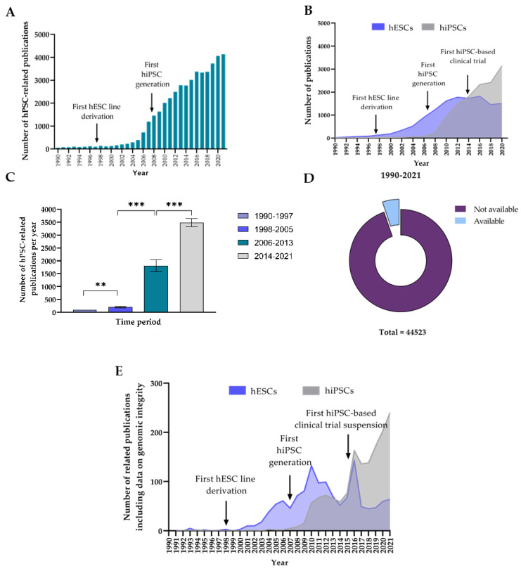
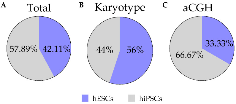
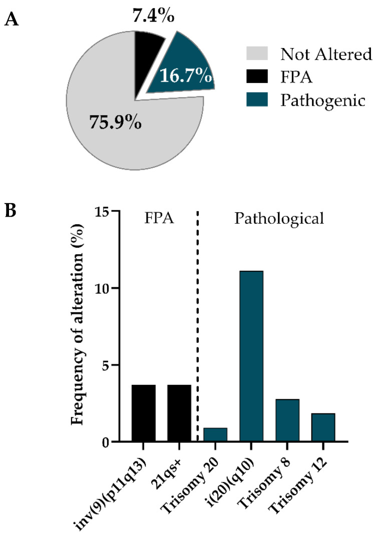
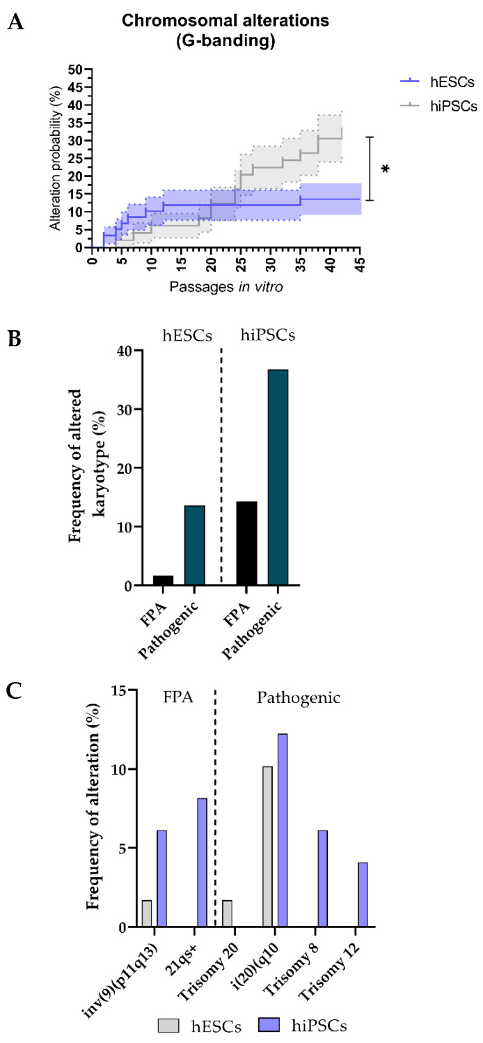
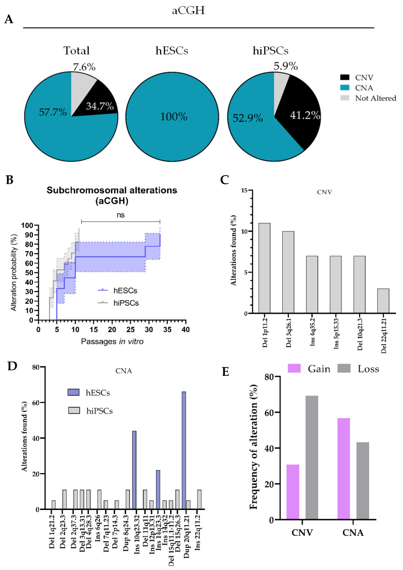
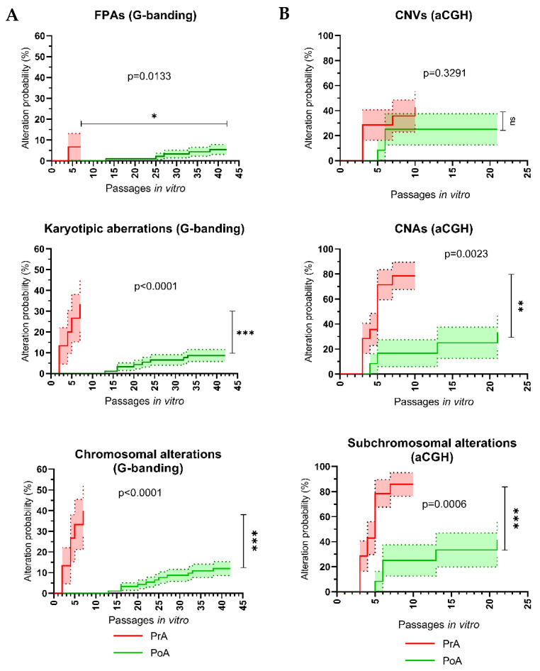
Human pluripotent stem cells (hPSCs) have generated unprecedented interest in the scientific community, given their potential applications in regenerative medicine, disease modeling, toxicology and drug screening. However, hPSCs are prone to acquire genomic alterations in vitro, mainly due to suboptimal culture conditions and inappropriate routines to monitor genome integrity. This poses a challenge to both the safety of clinical applications and the reliability of basic and translational hPSC research. In this study, we aim to investigate if the implementation of a Quality Management System (QMS) such as ISO9001:2015 to ensure reproducible and standardized cell culture conditions and genomic screening strategies can decrease the prevalence of genomic alterations affecting hPSCs used for research applications. To this aim, we performed a retrospective analysis of G-banding karyotype and Comparative Genomic Hybridization array (aCGH) data generated by our group over a 5-year span of different hESC and hiPSC cultures. This work demonstrates that application of a QMS to standardize cell culture conditions and genomic monitoring routines leads to a striking improvement of genomic stability in hPSCs cultured in vitro, as evidenced by a reduced probability of potentially pathogenic chromosomal aberrations and subchromosomal genomic alterations. These results support the need to implement QMS in academic laboratories performing hPSC research.

Keywords: GIVIMP; ISO9001; cell therapy; genomic instability; human pluripotent stem cells; karyotype.


Conflict of interest statement

The authors declare no conflict of interest. The funders had no role in the design of the study; in the collection, analyses, or interpretation of data; in the writing of the manuscript, or in the decision to publish the results.

Figures

Figure 1
Figure 1
Analysis of the evolution in number of human pluripotent stem cell (hPSC)-related publications and level of reporting of hPSC genomic integrity monitoring: (A) Bar graph representing the number of publications on hPSCs per year; (B) compound line graph of trend of publications per year during on human embryonic stem cells (hESCs) and human induced pluripotent stem cells (hiPSCs). Notice the three inflection points relative to three major events in the PSC field: the first hESC line derivation by Thomson in 1998 [2], the first hiPSC generation by Takahashi and Yamanaka in 2007 [4] and the RIKEN first hiPSC-based clinical trial in 2013; (C) histogram of average number of publications per year in distinct temporal windows; (D) publications of hPSC research containing information on genomic integrity monitoring of hPSCs. Available segment indicates the percentage of articles with published data about genomic integrity of hPSCs, where the mean ± the standard error of the mean (SEM) is showed; (E) trend of publications per year on hESCs and hiPSCs reporting information on genomic integrity of cells used for the study: increasing interest about monitoring genomic integrity follows critical events about their usage.
Figure 2
Figure 2
Descriptive statistics of the analyzed dataset. Pie charts showing the relative amount (%) of hESC and hiPSC lines used in this study (A), as well as the proportion of G-banding (B) and CGH array (aCGH) (C) which were performed in each of the hPSC types.
Figure 3
Figure 3
Recurrent karyotypic aberrations are detected in cultured hPSCs by G-banding: (A) Pie chart of fraction of genomic alterations detected with karyotype on the total of the cell lines analyzed; (B) histogram of chromosomal abnormalities found in hPSCs by using karyotype. Each bar represents the percentage of time that the alterations has been found in hPSCs. FPA: Frequent Polymorphic Alterations.
Figure 4
Figure 4
The hiPSCs lines show a higher prevalence of karyotype genomic aberrations detected by G-banding as compared to hESCs: (A) Survival analysis (Kaplan–Meier curves) of the evolution of the probability (%) of hESCs and hiPSCs being affected by chromosomal genomic alterations detected by G-banding over time (passages in vitro). Error bars represent standard error of the mean (s.e.m.); (B) bar graph of percentage of frequency of all alterations found in hESCs compared with hiPSCs on a total number of 108 karyotypes performed; (C) histogram showing the frequency at which different types of FPAs and pathologic alterations are detected in hESCs and hiPSCs via karyotype. FPA: Frequent Polymorphic Alterations.
Figure 5
Figure 5
Recurrent subchromosomal Copy Number Variations (CNVs) and pathogenic Copy Number Alterations (CNAs) are detected in cultured hESC s and hiPSCs by Comparative Genomic Hybridization CGH array (aCGH) analysis: (A) Pie charts showing the percentage of cells harboring Copy Number Variations (CNVs) or Copy Number Alterations (CNAs), as well as cell lines for which no alteration were detected; (B) survival analysis (Kaplan–Meier curves) of the evolution of the probability (%) of hESCs and hiPSCs being affected by subchromosomal genomic alterations detected by aCGH over time (passages in vitro). Error bars represent standard error (S.E.M.); (C,D) histograms showing the frequency at which different types of CNVs (C) and CNAs (D) are found in analyzed hESCs and hiPSCs; (E) bar graph showing the frequency at which DNA gains and losses are detected with aCGH for both CNV and CNA group.
Figure 6
Figure 6
Adaptation to standardized cell culture conditions described by standard operating procedures (SOPs) under a Quality Management System (QMS) results in improved genomic stability of cultured hPSCs. Survival analysis (Kaplan–Meier curves) of the evolution of the probability (%) of hPSCs being affected by de novo genomic alterations over time (passages in vitro) before and after adaptation to standardized cell culture conditions, using G-banding karyotyping (A) and Comparative Genome Hybridization array (aCGH; B) as detection methods. For each panel, the top, middle and bottom plots show the probability of hPSCs acquiring polymorphic alterations, pathogenic alterations and total alterations, respectively. Pre-adaptation (PrA) and post-adaptation (PoA) refer to hPSCs analyzed before and after completing adaptation to the standard cell culture conditions used in our laboratory, respectively. Error bars represent standard error (S.E.M.). FPA: Frequent Polymorphic Alteration; CNV: Copy Number Variation; can: Copy Number Alteration.

References

    1. Evans M.J., Kaufman M.H. Establishment in culture of pluripotential cells from mouse embryos. Nature. 1981;292:154–156. doi: 10.1038/292154a0. - DOI - PubMed
    1. Thomson J.A., Itskovitz-Eldor J., Shapiro S.S., Waknitz M.A., Swiergiel J.J., Marshall V.S., Jones J.M. Embryonic Stem Cell Lines Derived from Human Blastocysts. Science. 1998;282:1145–1147. doi: 10.1126/science.282.5391.1145. - DOI - PubMed
    1. Takahashi K., Yamanaka S. Induction of Pluripotent Stem Cells from Mouse Embryonic and Adult Fibroblast Cultures by Defined Factors. Cell. 2006;126:663–676. doi: 10.1016/j.cell.2006.07.024. - DOI - PubMed
    1. Takahashi K., Tanabe K., Ohnuki M., Narita M., Ichisaka T., Tomoda K., Yamanaka S. Induction of Pluripotent Stem Cells from Adult Human Fibroblasts by Defined Factors. Cell. 2007;131:861–872. doi: 10.1016/j.cell.2007.11.019. - DOI - PubMed
    1. Zhu Z., Huangfu D. Human pluripotent stem cells: An emerging model in developmental biology. Development. 2013;140:705–717. doi: 10.1242/dev.086165. - DOI - PMC - PubMed

Publication types