

Mechanotransduction in Skin Inflammation
- PMID: 35805110
- PMCID: PMC9265324
- DOI: 10.3390/cells11132026
Mechanotransduction in Skin Inflammation
Abstract
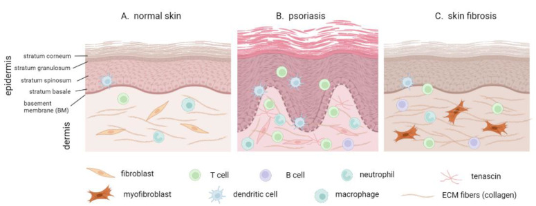
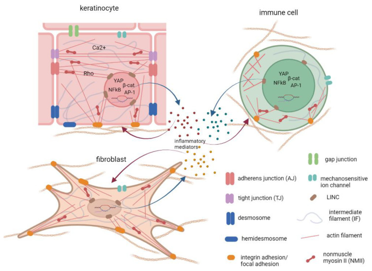
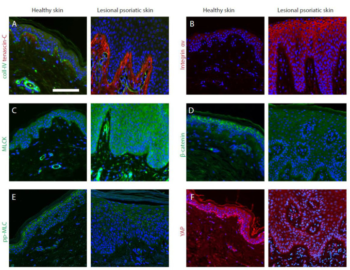
In the process of mechanotransduction, the cells in the body perceive and interpret mechanical stimuli to maintain tissue homeostasis and respond to the environmental changes. Increasing evidence points towards dysregulated mechanotransduction as a pathologically relevant factor in human diseases, including inflammatory conditions. Skin is the organ that constantly undergoes considerable mechanical stresses, and the ability of mechanical factors to provoke inflammatory processes in the skin has long been known, with the Koebner phenomenon being an example. However, the molecular mechanisms and key factors linking mechanotransduction and cutaneous inflammation remain understudied. In this review, we outline the key players in the tissue's mechanical homeostasis, the available data, and the gaps in our current understanding of their aberrant regulation in chronic cutaneous inflammation. We mainly focus on psoriasis as one of the most studied skin inflammatory diseases; we also discuss mechanotransduction in the context of skin fibrosis as a result of chronic inflammation. Even though the role of mechanotransduction in inflammation of the simple epithelia of internal organs is being actively studied, we conclude that the mechanoregulation in the stratified epidermis of the skin requires more attention in future translational research.
Keywords: actin-myosin cytoskeleton; atopic dermatitis; cytokines; epidermis; fibrosis; integrins; intermediate filaments; keratinocyte; psoriasis; stretch.
Conflict of interest statement
The authors declare no conflict of interest.
Figures
Similar articles
-
Keratinocyte Expression of A20/TNFAIP3 Controls Skin Inflammation Associated with Atopic Dermatitis and Psoriasis.J Invest Dermatol. 2019 Jan;139(1):135-145. doi: 10.1016/j.jid.2018.06.191. Epub 2018 Aug 14. J Invest Dermatol. 2019. PMID: 30118730
-
Increase in primary cilia in the epidermis of patients with atopic dermatitis and psoriasis.Exp Dermatol. 2021 Jun;30(6):792-803. doi: 10.1111/exd.14285. Epub 2021 Feb 12. Exp Dermatol. 2021. PMID: 33455013
-
Annoying Psoriasis and Atopic Dermatitis: A Narrative Review.Int J Mol Sci. 2022 Apr 28;23(9):4898. doi: 10.3390/ijms23094898. Int J Mol Sci. 2022. PMID: 35563285 Free PMC article. Review.
-
Emerging Role of the IL-36/IL-36R Axis in Multiple Inflammatory Skin Diseases.J Invest Dermatol. 2024 Feb;144(2):206-224. doi: 10.1016/j.jid.2023.11.004. Epub 2024 Jan 7. J Invest Dermatol. 2024. PMID: 38189700 Review.
-
Unraveling the Role of Sex Hormones on Keratinocyte Functions in Human Inflammatory Skin Diseases.Int J Mol Sci. 2022 Mar 15;23(6):3132. doi: 10.3390/ijms23063132. Int J Mol Sci. 2022. PMID: 35328552 Free PMC article. Review.
Cited by
-
Evidence Supporting Conservative Scar Management Interventions Following Burn Injury: A Review Article.J Burn Care Res. 2025 Aug 12;46(3):504-514. doi: 10.1093/jbcr/irae204. J Burn Care Res. 2025. PMID: 39548761 Free PMC article. Review.
-
Therapeutic Effect of Liquiritin Carbomer Gel on Topical Glucocorticoid-Induced Skin Inflammation in Mice.Pharmaceutics. 2024 Jul 28;16(8):1001. doi: 10.3390/pharmaceutics16081001. Pharmaceutics. 2024. PMID: 39204346 Free PMC article.
-
Hydrogel dressing integrating FAK inhibition and ROS scavenging for mechano-chemical treatment of atopic dermatitis.Nat Commun. 2023 Apr 29;14(1):2478. doi: 10.1038/s41467-023-38209-x. Nat Commun. 2023. PMID: 37120459 Free PMC article.
-
The mechanotransducer Piezo1 coordinates metabolism and inflammation to promote skin growth.Nat Commun. 2025 Jul 25;16(1):6876. doi: 10.1038/s41467-025-62270-3. Nat Commun. 2025. PMID: 40715139 Free PMC article.
-
Bioinspired and Photo-Clickable Thiol-Ene Bioinks for the Extrusion Bioprinting of Mechanically Tunable 3D Skin Models.Biomimetics (Basel). 2024 Apr 10;9(4):228. doi: 10.3390/biomimetics9040228. Biomimetics (Basel). 2024. PMID: 38667239 Free PMC article.
References
-
- Moll I. Duale Reihe Dermatologie. Thieme; Stuttgart, Deutschland: 2016.
Publication types
MeSH terms
LinkOut - more resources
Full Text Sources
Medical
