Review
. 2022 Jun 29;11(13):2067.
doi: 10.3390/cells11132067.

Insights into the Structure and Function of the Pex1/Pex6 AAA-ATPase in Peroxisome Homeostasis

Affiliations
Review

Insights into the Structure and Function of the Pex1/Pex6 AAA-ATPase in Peroxisome Homeostasis

Ryan M Judy et al. Cells. .

Abstract

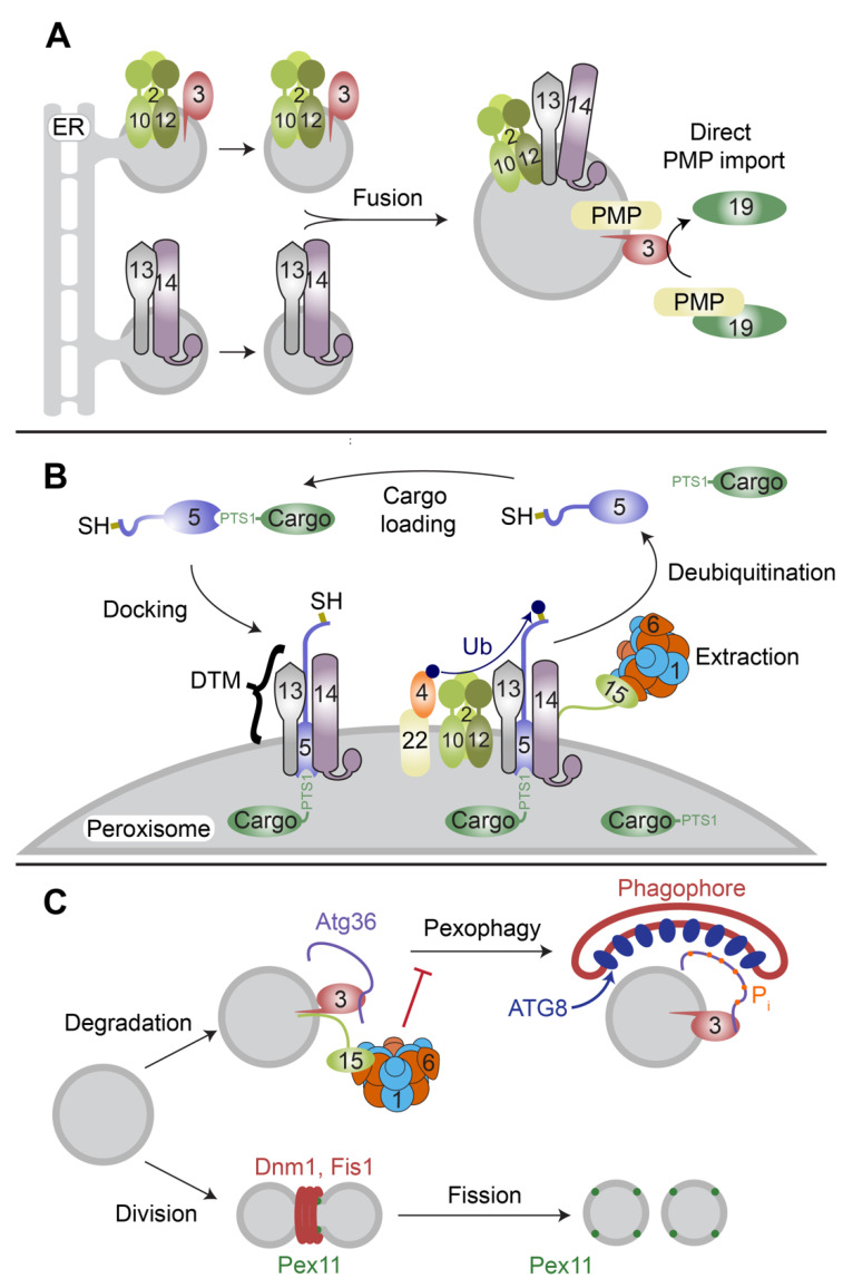
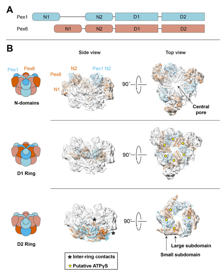
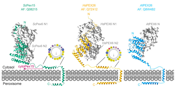
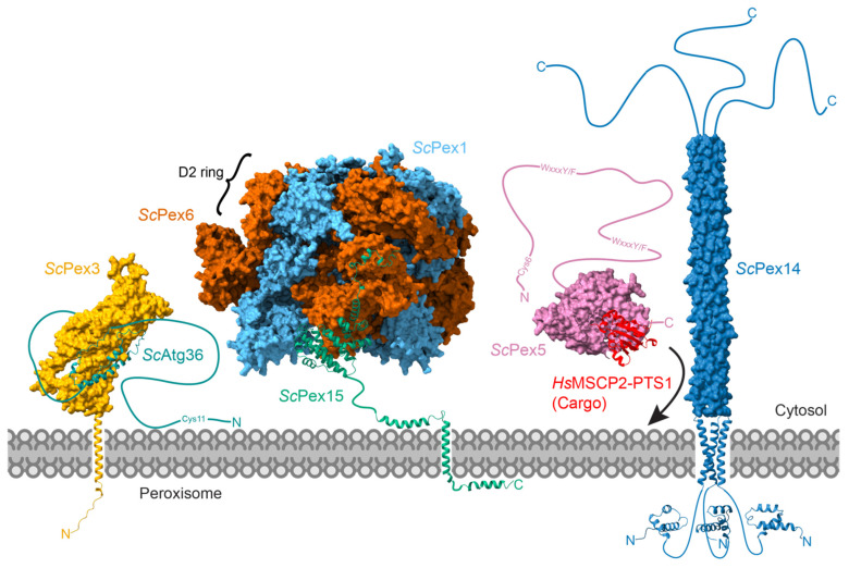
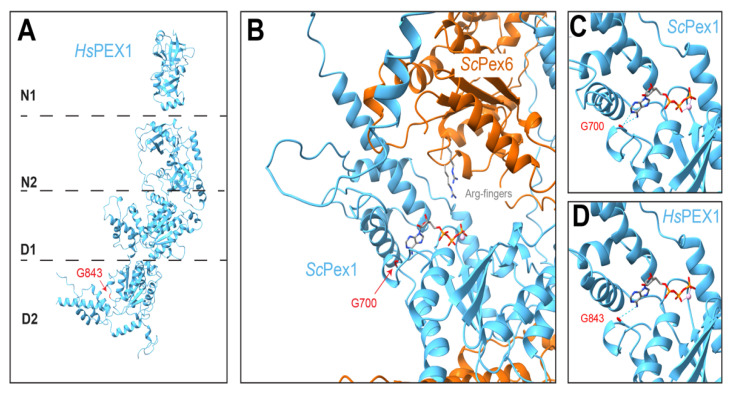
The AAA-ATPases Pex1 and Pex6 are required for the formation and maintenance of peroxisomes, membrane-bound organelles that harbor enzymes for specialized metabolism. Together, Pex1 and Pex6 form a heterohexameric AAA-ATPase capable of unfolding substrate proteins via processive threading through a central pore. Here, we review the proposed roles for Pex1/Pex6 in peroxisome biogenesis and degradation, discussing how the unfolding of potential substrates contributes to peroxisome homeostasis. We also consider how advances in cryo-EM, computational structure prediction, and mechanisms of related ATPases are improving our understanding of how Pex1/Pex6 converts ATP hydrolysis into mechanical force. Since mutations in PEX1 and PEX6 cause the majority of known cases of peroxisome biogenesis disorders such as Zellweger syndrome, insights into Pex1/Pex6 structure and function are important for understanding peroxisomes in human health and disease.

Keywords: AAA-ATPase; PEX1; PEX26; PEX6; organelle biogenesis; peroxisomes; translocation.


Conflict of interest statement

The authors declare no conflict of interest.

Figures

Figure 1
Figure 1
Overview of S. cerevisiae peroxisome biogenesis. (A) In de novo biogenesis, pre-peroxisomal vesicles (ppVs) carrying peroxisome membrane proteins bud and fuse with other ppVs or with mature peroxisomes. PMPs can also be directly inserted into the peroxisomal membrane by Pex19 and Pex3. (B) Pex5 binds the C-terminal PTS1-targeting signal on a cargo protein and interacts with the docking and translocation module (DTM) to import the cargo protein into the peroxisome. Pex5 is subsequently ubiquitinated by Pex4 and the Pex2/Pex10/Pex12 complex. Pex1/Pex6 extracts Pex5 from the peroxisomal membrane. Following deubiquitination, Pex5 can repeat the import cycle. (C) Mature peroxisomes can either grow and divide using the peroxisomal fission factor Pex11 and shared mitochondrial factors (Dnm1 and Fis1) or undergo peroxisome-specific autophagy mediated by adaptors such as Atg36. Pex1/Pex6 interacts with Atg36 through Pex3 and Pex15 to suppress Atg36 phosphorylation and pexophagy. Accessory proteins involved in process are represented as numbered proteins.
Figure 2
Figure 2
Pex1/Pex6 is a double ring hexameric complex with alternating subunits. (A) Pex1 and Pex6 each have two N-terminal domains and two ATPase domains. Pex1 N1 is flexibly attached to the rest of the complex. (B) S. cerevisiae Pex1/Pex6 structure, based on AlphaFold2 models (red and blue) split by the domain and fitted into EMDB-6359 (gray) [143]. Map resolution: 7.2 Å; fit correlation coefficient: 0.83. The Pex1 and Pex6 N2 domains bind above the D1 ATPase ring, while the Pex6 N1 domain binds to the side of the D1 ATPase ring. The Pex1 N1 domain was not resolved. The D1 ATPase ring binds but does not hydrolyze ATP and is thought to contribute to hexamer assembly. The top view of the active D2 ATPase ring is displayed at a lower threshold than other maps. Asterisks show sites of contact between D1 and D2 rings (see text). Stars represent expected ATP-binding sites based on inter-protomer distances [143].
Figure 3
Figure 3
(A) The Alphafold2 predictions for Pex1 N1 domain structures in human (yellow), S. cerevisiae (green), and A. thaliana (blue) aligned to the X-ray crystal structure of murine PEX1 N1 (gray, PDB: 1WLF). The yellow/red ligand represents a sulfate from the PEX1 N1 crystal structure. (B) Sequence conservation mapped on the human PEX1 N1 domain. Cofactors such as Npl4 and FAF1 bind a similar cleft between subdomains (arrow) in Cdc48-N. (C) Coulombic potential mapped on the surface of human PEX1 N1 domain showing negative charge in the conserved region.
Figure 4
Figure 4
Structures of Pex1 (blue) and Pex6 (orange) monomers from AlphaFold2 or, for HsPEX1 N1, X-ray crystallography (PDB 1WLF, [148]). N1 domains are separated for clarity; Pex1 N1 domains are flexibly attached to the motor, while Pex6 N1 domains are rigidly attached to the Pex6 D1 ring (see Figure 2).
Figure 5
Figure 5
Structural models for the Pex1/Pex6 peroxisomal tethers: S. cerevisiae Pex15, H. sapiens PEX26, and A. thalania PEX26, based AlphaFold2 multimer predictions. Pathogenic missense mutations in PEX26 (D43H, L44P, L45P, G89R, R98W, P117L, and P118R) are colored in blue (Leiden Open Variation Database 3.0, [199]). Predictions for transmembrane domains are from the highest scores from TMHMM-2.0 [200]. Helical wheels generated in Heliquest [201], letters indicate predicted protein sequence.
Figure 6
Figure 6
Putative Pex1/Pex6 substrates (cytoPex15, Pex5, and Atg36) have disordered tails (curved lines) capable of accessing the Pex1/Pex6 D2 pore. Pex1/Pex6 is recruited to the peroxisome membrane by Pex15 (models based on EMDB-6359; PDB 5VXV; Alphafold2 multimer). In vitro, the truncated cytosolic domain of Pex15 is a Pex1/Pex6 substrate. Pex5 bound to a PTS1-tagged protein (model based on HsPEX5 bound to MSCP2 PDB: 2C0L; Alphafold2 ScPex5 AF: P35056) embeds in the DTM, primarily composed of Pex5 bound to Pex14 (modeled from EMDB-12047 [10.2 Å]; Alphafold2 AF: P53122). Pex1/Pex6 is then thought to extract mono-ubiquitinated Pex5 from the membrane. Atg36 interacts with Pex1/Pex6 indirectly through Pex3 and Pex15 (models based on Alphafold2 multimer). Pex1/Pex6 prevents Atg36 phosphorylation, though the mechanism is unclear. Figures made with ChimeraX 1.3 [147].
Figure 7
Figure 7
PEX1 G843D is expected to disrupt ATP binding and/or protomer folding. (A) HsPEX1, based on AlphaFold2 and X-ray crystallography (as in Figure 2, PDB 1WLF, [148]). G843 is in the PEX1 D2 ring. (B) ScPex1 D2 ATPase in the Pex1/Pex6 hexamer at the Pex1 site most likely to be nucleotide-bound (AlphaFold2, EMDB-6359, [143]). Note that nucleotides are not discernible in experimental structures of Pex1/Pex6; an ATP is modeled based on alignment with a high-resolution structure of Hsp97 (PDB 7LN5, [159]). (C) The glycine G843 in HsPEX1 is conserved. The homologous residue in ScPex1 (G700) is colored in red and the backbone is predicted to hydrogen bond with the adenosine of ATP ([159], ChimeraX). (D) In the structurally similar ScPex1 and HsPEX1 ATPase sites, G700 or G843 hydrogen bond (dotted lines) with ATP.

Similar articles

Cited by

References

    1. Gabaldón T. Peroxisome Diversity and Evolution. Philos. Trans. R. Soc. B Biol. Sci. 2010;365:765–773. doi: 10.1098/rstb.2009.0240. - DOI - PMC - PubMed
    1. Jansen R.L.M., Santana Molina C., Van Den Noort M., Devos D.P., Van Der Klei I.J. Comparative Genomics of Peroxisome Biogenesis Proteins: Making Sense of the PEX Proteins. Front. Cell Dev. Biol. 2021;9:654163. doi: 10.3389/fcell.2021.654163. - DOI - PMC - PubMed
    1. Nordgren M., Fransen M. Peroxisomal Metabolism and Oxidative Stress. Biochimie. 2014;98:56–62. doi: 10.1016/j.biochi.2013.07.026. - DOI - PubMed
    1. Goldman B.M., Blobel G. Biogenesis of Peroxisomes: Intracellular Site of Synthesis of Catalase and Uricase. Proc. Natl. Acad. Sci. USA. 1978;75:5066–5070. doi: 10.1073/pnas.75.10.5066. - DOI - PMC - PubMed
    1. Gurvitz A., Rottensteiner H. The Biochemistry of Oleate Induction: Transcriptional Upregulation and Peroxisome Proliferation. Biochim. Biophys. Acta BBA-Mol. Cell Res. 2006;1763:1392–1402. doi: 10.1016/j.bbamcr.2006.07.011. - DOI - PubMed

Publication types

MeSH terms

Substances