Review
. 2022 Jun 30;11(13):2095.
doi: 10.3390/cells11132095.

Regeneration or Repair? The Role of Alveolar Epithelial Cells in the Pathogenesis of Idiopathic Pulmonary Fibrosis (IPF)

Affiliations
Review

Regeneration or Repair? The Role of Alveolar Epithelial Cells in the Pathogenesis of Idiopathic Pulmonary Fibrosis (IPF)

Paola Confalonieri et al. Cells. .

Abstract

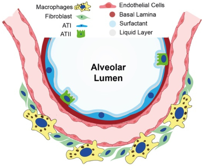
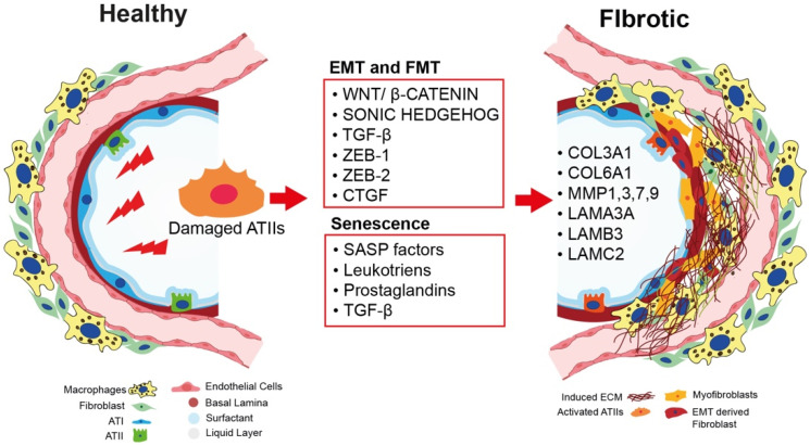
Idiopathic pulmonary fibrosis (IPF) is a chronic, progressive interstitial lung disease (ILD) with unknown etiology in which gradual fibrotic scarring of the lungs leads to usual interstitial pneumonia (UIP) and, ultimately, to death. IPF affects three million people worldwide, and the only currently available treatments include the antifibrotic drugs nintedanib and pirfenidone, which effectively reduce fibrosis progression are, unfortunately, not effective in curing the disease. In recent years, the paradigm of IPF pathogenesis has shifted from a fibroblast-driven disease to an epithelium-driven disease, wherein, upon recurrent microinjuries, dysfunctional alveolar type II epithelial cells (ATII) are not only unable to sustain physiological lung regeneration but also promote aberrant epithelial-mesenchymal crosstalk. This creates a drift towards fibrosis rather than regeneration. In the context of this review article, we discuss the most relevant mechanisms involved in IPF pathogenesis with a specific focus on the role of dysfunctional ATII cells in promoting disease progression. In particular, we summarize the main causes of ATII cell dysfunction, such as aging, environmental factors, and genetic determinants. Next, we describe the known mechanisms of physiological lung regeneration by drawing a parallel between embryonic lung development and the known pathways involved in ATII-driven alveolar re-epithelization after injury. Finally, we review the most relevant interventional clinical trials performed in the last 20 years with the aim of underlining the urgency of developing new therapies against IPF that are not only aimed at reducing disease progression by hampering ECM deposition but also boost the physiological processes of ATII-driven alveolar regeneration.

Keywords: ATII cells; IPF; regeneration; senescence.


Conflict of interest statement

The authors declare no conflict of interest.

Figures

Figure 1
Figure 1
Cell composition of the healthy alveolus. Schematical image of the alveolar compartment: the alveolar lumen (white) is surrounded by ATI cells (blue) and ATII cells (green), which secrete surfactant (light blue). The alveolar epithelial basal lamina (pink) separates alveolar epithelial cells from alveolar endothelial cells. Pulmonary interstitial tissue is schematically represented as a compartment mainly composed of fibroblasts (light green) and macrophages (yellow), which are known to be the most relevant interstitial cell types in the pathogenesis of IPF.
Figure 2
Figure 2
Key processes inducing pulmonary fibrosis. Repetitive injuries in human adult lung lead to damaged ATII cells (orange). Damaged ATII cells begin overexpressing the Wnt/β-catenin and SHH signaling pathways and secreting TGF-β. They also start expressing ZEB-1, ZEB-2, and CTGF as a response to TGF-β secretion. These factors are involved in the EMT and FMT processes. In parallel, damaged ATII cells acquire a senescent phenotype, producing SASP factors, leukotrienes, and prostaglandins. These factors collectively contribute to the proliferation of myofibroblasts which, in turn, are responsible for the aberrant deposition of ECM components, such as COL3A1, COL6A1, MMP1, MMP3, MMP7, MMP9, LAMA3, LAMB3, and LAMC2.
Figure 3
Figure 3
Transdifferentiation process from progenitor cells to ATI cells. Schematic depiction of the sequential differentiation process that occurs during cell differentiation into ATI cells as a response to injury, starting from a niche of SOX9/SOX2/NKX2.1-positive progenitor cells, which differentiate into ATII cells expressing SPC, SPA, and ABCA3. ATII cells then undergo a transition phase, where they begin to express KRT8 and markers of both ATII (SPC) and ATI cells (RAGE). Finally, from this intermediate state, cells transdifferentiate into terminally differentiated ATI cells positive for RAGE, podoplanin (PDPLN), and aquaporin 5 (AQP5).
Figure 4
Figure 4
Novel therapeutic strategies for IPF treatment. Summary illustration of putative strategies to (1) stop fibrosis progression by hampering aberrant ECM deposition and eliminating defective cells (antifibrotic and senolytic drugs) and (2) bring back the lung to a healthy condition by promoting alveolar regeneration (regenerative therapies).

References

    1. Navaratnam V., Fleming K.M., West J., Smith C.J., Jenkins R.G., Fogarty A., Hubbard R.B. The rising incidence of idiopathic pulmonary fibrosis in the U.K. Thorax. 2011;66:462–467. doi: 10.1136/thx.2010.148031. - DOI - PubMed
    1. King T.E., Jr., Pardo A., Selman M. Idiopathic pulmonary fibrosis. Lancet. 2011;378:1949–1961. doi: 10.1016/S0140-6736(11)60052-4. - DOI - PubMed
    1. Ley B., Collard H.R. Epidemiology of idiopathic pulmonary fibrosis. Clin. Epidemiol. 2013;5:483–492. doi: 10.2147/CLEP.S54815. - DOI - PMC - PubMed
    1. Travis W.D., Costabel U., Hansell D.M., King T.E., Jr., Lynch D.A., Nicholson A.G., Ryerson C.J., Ryu J.H., Selman M., Wells A.U., et al. An official American Thoracic Society/European Respiratory Society statement: Update of the international multidisciplinary classification of the idiopathic interstitial pneumonias. Am. J. Respir. Crit. Care Med. 2013;188:733–748. doi: 10.1164/rccm.201308-1483ST. - DOI - PMC - PubMed
    1. Collard H.R., Chen S.Y., Yeh W.S., Li Q., Lee Y.C., Wang A., Raghu G. Health care utilization and costs of idiopathic pulmonary fibrosis in U.S. Medicare beneficiaries aged 65 years and older. Ann. Am. Thorac. Soc. 2015;12:981–987. doi: 10.1513/AnnalsATS.201412-553OC. - DOI - PubMed

LinkOut - more resources