. 2022 Jun 22;23(13):6927.
doi: 10.3390/ijms23136927.

Bone Tissue Engineering in Rat Calvarial Defects Using Induced Bone-like Tissue by rhBMPs from Immature Muscular Tissues In Vitro

Affiliations

Bone Tissue Engineering in Rat Calvarial Defects Using Induced Bone-like Tissue by rhBMPs from Immature Muscular Tissues In Vitro

Tatsuhide Hayashi et al. Int J Mol Sci. .

Abstract

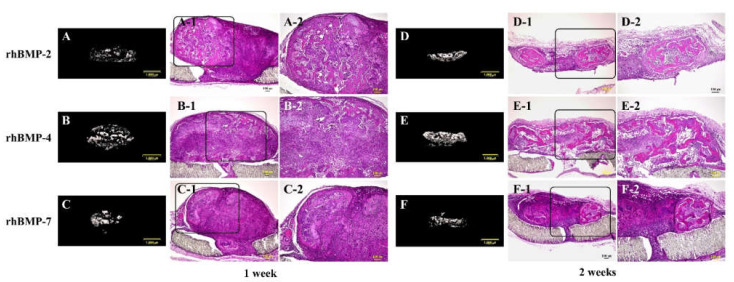
This study aimed to induce bone-like tissue from immature muscular tissue (IMT) in vitro using commercially available recombinant human bone morphogenetic protein (rhBMP)-2, rhBMP-4, and rhBMP-7, and then implanting this tissue into a calvarial defect in rats to assess healing. IMTs were extracted from 20-day-old Sprague-Dawley (SD) fetal rats, placed on expanded polytetrafluoroethylene (ePTFE) with 10 ng/μL each of rhBMP-2, BMP-4, and BMP-7, and cultured for two weeks. The specimens were implanted into calvarial defects in 3-week-old SD rats for up to three weeks. Relatively strong radiopacity was observed on micro-CT two weeks after culture, and bone-like tissue, comprising osteoblastic cells and osteoids, was partially observed by H&E staining. Calcium, phosphorus, and oxygen were detected in the extracellular matrix using an electron probe micro analyzer, and X-ray diffraction patterns and Fourier transform infrared spectroscopy spectra of the specimen were found to have typical apatite crystal peaks and spectra, respectively. Furthermore, partial strong radiopacity and ossification were confirmed one week after implantation, and a dominant novel bone was observed after two weeks in the defect site. Thus, rhBMP-2, BMP-4, and BMP-7 differentiated IMT into bone-like tissue in vitro, and this induced bone-like tissue has ossification potential and promotes the healing of calvarial defects. Our results suggest that IMT is an effective tissue source for bone tissue engineering.

Keywords: bone tissue engineering; expanded polytetrafluoroethylene; immature muscular tissue; induced bone-like tissue; recombinant human bone morphogenetic proteins.


Conflict of interest statement

The authors declare no conflict of interest.

Figures

Figure 1
Figure 1
Typical micro-CT (AD) and histological images of cultured immature muscular tissue (IMT), control (treated with 10 μL of sterilized 4 mM HCl containing 0.2% BSA only) and treated with 10 μL of each recombinant human bone morphogenetic protein (rhBMP)-2, rhBMP-4, and rhBMP-7. Samples were cultured for two weeks. A-1, -2, B-1, -2, C-1, -2, and D-1, -2 show hematoxylin and eosin (H&E) staining and A-3, -4, B-3, -4, C-3, -4, and D-3, -4 show von Kossa staining. A-2–D-2 and A-4–D-4 are magnified insert images of A-1–D-1 and A-3–D-3, respectively. Scale bar sizes in micro-CT and histological images are 1000 µm and 100 µm, respectively.
Figure 2
Figure 2
EPMA analysis of the elemental compositions of rhBMP-2, rhBMP-4, and rhBMP-7 samples two weeks after cultivation. Calcium (Ca; red), phosphorus (P; green), and oxygen (O; blue) were detected in the extracellular matrix. All three elements were confirmed to be at almost identical positions (white). Scale bar size is 10 µm.
Figure 3
Figure 3
XRD patterns of rhBMP-2, rhBMP-4, and rhBMP-7 samples two weeks after cultivation and rat calvarial cortical bone. Black triangles show typical apatite peaks and their crystal planes were indexed according to JCPDS card 9-432.
Figure 4
Figure 4
FT-IR spectra of rhBMP-2, rhBMP-4, and rhBMP-7 samples two weeks after cultivation and rat calvarial cortical bone.
Figure 5
Figure 5
Typical micro-CT (AC and DF) and histological images of each subcutaneously implanted rhBMP-2, rhBMP-4, and rhBMP-7 sample into the backs of rats at one and two weeks. A-2, B-2, and C-2 are magnified insert images of A-1, B-1, and C-1, and D-2, E-2, and F-2 are magnified insert images of D-1, E-1, and F-1, respectively. Scale bar sizes in micro-CT and histological images are 1000 µm and 100 µm, respectively.
Figure 6
Figure 6
Typical micro-CT (AD, EH, and IL) and histological images of each implanted rhBMP-2, rhBMP-4, and rhBMP-7 sample into the calvarial defects of rats at one, two, and three weeks. (A,E,I) are the control. A-2–D-2, E-2–H-2, and I-2–L-2 are magnified insert images in A-1–D-1, E-1–H-1, and I-1–L-1, respectively. The circles in J-2 and K-2 indicate the areas where the induced new bone integrated with the original calvarial bone. Comparison of the new bone volume of defect area (%) at one (M), two (N), and three (O) weeks after implantation. The data are shown as mean ± SD (n = 3). Scale bar sizes in micro-CT and histological images are 2000 µm and 100 µm, respectively.

Similar articles

Cited by

References

    1. Roddy E., DeBaun M.R., Daoud-Gray A., Yang Y.P., Gardner M.J. Treatment of critical-sized bone defects: Clinical and tissue engineering perspectives. Eur. J. Orthop. Surg. Traumatol. 2018;28:351–362. doi: 10.1007/s00590-017-2063-0. - DOI - PubMed
    1. Oryan A., Alidadi S., Moshiri A., Maffulli N. Bone regenerative medicine: Classic options, novel strategies, and future directions. J. Orthop. Surg. Res. 2014;9:18. doi: 10.1186/1749-799X-9-18. - DOI - PMC - PubMed
    1. Langer R., Vacanti J.P. Tissue engineering. Science. 1993;260:920–926. doi: 10.1126/science.8493529. - DOI - PubMed
    1. Tabata Y. Tissue regeneration based on tissue engineering technology. Congenit. Anom. 2004;44:111–124. doi: 10.1111/j.1741-4520.2004.00024.x. - DOI - PubMed
    1. Langer R., Vacanti J. Advances in tissue engineering. J. Pediatr. Surg. 2016;51:8–12. doi: 10.1016/j.jpedsurg.2015.10.022. - DOI - PMC - PubMed

Substances

LinkOut - more resources