Review
. 2022 Jun 22;23(13):6937.
doi: 10.3390/ijms23136937.

Roles of ATP and SERCA in the Regulation of Calcium Turnover in Unloaded Skeletal Muscles: Current View and Future Directions

Affiliations
Review

Roles of ATP and SERCA in the Regulation of Calcium Turnover in Unloaded Skeletal Muscles: Current View and Future Directions

Tatiana L Nemirovskaya et al. Int J Mol Sci. .

Abstract

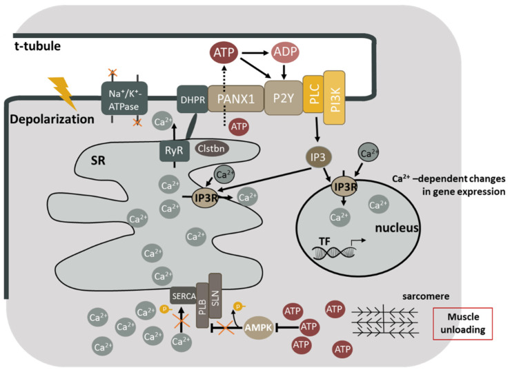
A decrease in skeletal muscle contractile activity or its complete cessation (muscle unloading or disuse) leads to muscle fibers' atrophy and to alterations in muscle performance. These changes negatively affect the quality of life of people who, for one reason or another, are forced to face a limitation of physical activity. One of the key regulatory events leading to the muscle disuse-induced changes is an impairment of calcium homeostasis, which leads to the excessive accumulation of calcium ions in the sarcoplasm. This review aimed to analyze the triggering mechanisms of calcium homeostasis impairment (including those associated with the accumulation of high-energy phosphates) under various types of muscle unloading. Here we proposed a hypothesis about the regulatory mechanisms of SERCA and IP3 receptors activity during muscle unloading, and about the contribution of these mechanisms to the excessive calcium ion myoplasmic accumulation and gene transcription regulation via excitation-transcription coupling.

Keywords: SERCA; calcium signaling; modeled microgravity; muscle disuse; muscle unloading.


Conflict of interest statement

The authors declare no conflict of interest.

Figures

Figure 1
Figure 1
Molecular mechanisms contributing to the sarcoplasmic calcium accumulation during skeletal muscle unloading. Inactivation of Na,K-ATPase leads to sarcolemma depolarization, which in turn causes the activation of DHPR and the opening of RyR. Sarcoplasmic calcium accumulation leads to the detachment of calstabin from the ryanodine receptor and the occurrence of calcium leakage. Meanwhile, the inactivation of AMP-dependent protein kinase leads to dephosphorylation of PLN, and the blockage of SERCA. Excessive accumulation of calcium ions in the sarcoplasm causes calcium-dependent proteolysis and changes in gene expression in muscle fibers. ATP is released by PANX1 under functional muscle unloading. ATP is rapidly degraded to ADP, AMP, and adenosine under the action of ectonucleotidases. ATP and ADP can affect P2Y receptors associated with G proteins that in turn activate PI3 kinase. PI3 kinase catalyzes phosphorylation of phosphatidylinositol diphosphate (PIP2), giving PIP3 a highly charged residue, which recruits phospholipase C into the membrane, triggering the formation of inositol-1,4,5-triphosphate (IP3). IP3 then binds to IP3 receptors (IP3R) present both in the nuclear envelope and in the sarcoplasmic network, causing a weak signal of calcium release both in the cytosol and in the nucleoplasm, which contributes (probably, with other signaling cascades) to the activation of transcription factors (TF) leading to the expression or repression of the genes involved in the phenotype of muscle cells. DHPR—dihydropyridine channels; RyR—ryanodine receptors; SERCA—sarcoplasmic calcium-dependent ATPase; Clstbn—calstabin; AMPK—AMP-dependent protein kinase; PANX1—pannexin channels; P2Y—P2Y receptors; G—g-protein; PI3K—PI3-kinase; IP3R—IP3 receptors (IP3R—inositol 1,4,5-triphosphate receptors); IP3—inositol 1,4,5-triphosphate; PLC—phospholipase C.

References

    1. Shenkman B.S. How Postural Muscle Senses Disuse? Early Signs and Signals. Int. J. Mol. Sci. 2020;21:5037. doi: 10.3390/ijms21145037. - DOI - PMC - PubMed
    1. Belova S.T., Tyganov S.A., Mochalova E., Shenkman B. Restricted Activity and Protein Synthesis in Postural and Locomotor Muscles. Ross. Fiziol. Zh. Im. I.M. Sechenova. 2021;107:842–853. doi: 10.1134/S0022093021030194. - DOI
    1. Monti E., Reggiani C., Franchi M.V., Toniolo L., Sandri M., Armani A., Zampieri S., Giacomello E., Sarto F., Sirago G., et al. Neuromuscular junction instability and altered intracellular calcium handling as early determinants of force loss during unloading in humans. J. Physiol. 2021;599:3037–3061. doi: 10.1113/JP281365. - DOI - PMC - PubMed
    1. Georgiev T., Svirin M., Jaimovich E., Fink R.H. Localized nuclear and perinuclear Ca2+ signals in intact mouse skeletal muscle fibers. Front. Physiol. 2015;6:263. doi: 10.3389/fphys.2015.00263. - DOI - PMC - PubMed
    1. Taylor C.W., Tovey S.C. IP(3) receptors: Toward understanding their activation. Cold Spring Harb. Perspect. Biol. 2010;2:a004010. doi: 10.1101/cshperspect.a004010. - DOI - PMC - PubMed

LinkOut - more resources