. 2022 Jun 23;23(13):6961.
doi: 10.3390/ijms23136961.

Lmx1a-Dependent Activation of miR-204/211 Controls the Timing of Nurr1-Mediated Dopaminergic Differentiation

Affiliations

Lmx1a-Dependent Activation of miR-204/211 Controls the Timing of Nurr1-Mediated Dopaminergic Differentiation

Salvatore Pulcrano et al. Int J Mol Sci. .

Abstract

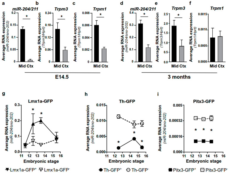
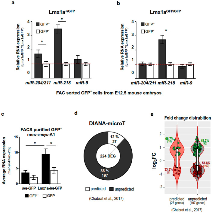
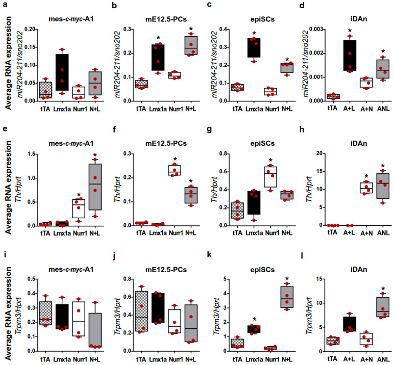
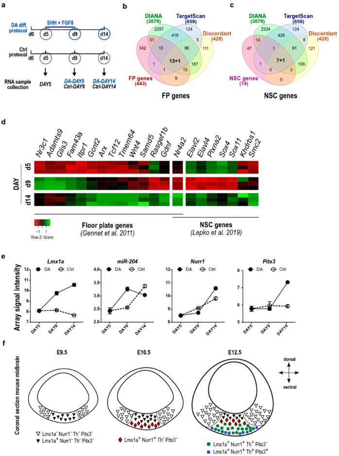
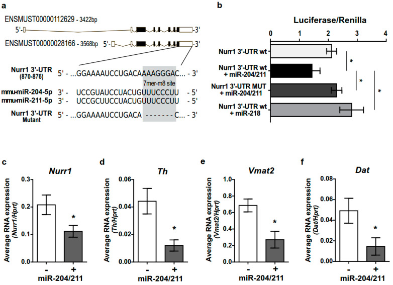
The development of midbrain dopaminergic (DA) neurons requires a fine temporal and spatial regulation of a very specific gene expression program. Here, we report that during mouse brain development, the microRNA (miR-) 204/211 is present at a high level in a subset of DA precursors expressing the transcription factor Lmx1a, an early determinant for DA-commitment, but not in more mature neurons expressing Th or Pitx3. By combining different in vitro model systems of DA differentiation, we show that the levels of Lmx1a influence the expression of miR-204/211. Using published transcriptomic data, we found a significant enrichment of miR-204/211 target genes in midbrain dopaminergic neurons where Lmx1a was selectively deleted at embryonic stages. We further demonstrated that miR-204/211 controls the timing of the DA differentiation by directly downregulating the expression of Nurr1, a late DA differentiation master gene. Thus, our data indicate the Lmx1a-miR-204/211-Nurr1 axis as a key component in the cascade of events that ultimately lead to mature midbrain dopaminergic neurons differentiation and point to miR-204/211 as the molecular switch regulating the timing of Nurr1 expression.

Keywords: Lmx1a; Nurr1; dopamine; microRNA.


Conflict of interest statement

The authors declare no conflict of interest.

Figures

Figure 1
Figure 1
miR-204/211 is enriched in a DA precursor subpopulation during midbrain development. (af) TaqMan assay for miR-204/211 (a,d) and qPCR for Trpm3 (b,e) and Trpm1 (c,f) on microdissected midbrain (Mid) and cortex (Ctx) at E14.5 (ac) or 3 months (df). miR-204/211 values are normalized to sno-202 expression, while mRNA levels are normalized on the reference mRNA hypoxanthine phosphoribosyl transferase (Hprt). Bars represent mean ± SD of 2−ΔCt values from four animals. * p < 0.05 (unpaired t-test with Welch’s correction). (gi) TaqMan assay for the expression of miR-204/211 in FACS-purified GFP+ and GFP cells from Lmx1a-GFP (g), Th-GFP (h), and Pitx3-GFP (i) reporter mice at different developmental stages. Data are normalized to the average of the reference sno-202 and represent the mean ± SD of 2−ΔCt values from three independent experiments. * p < 0.05 of GFP+ with respect to GFP (two-way ANOVA followed by Sidak).
Figure 2
Figure 2
Lmx1a levels influence miR-204/211 expression and its repressive activity. (a,b) TaqMan assay for miR-204/211, miR-218, and miR-9 on FACS-purified GFP+ and GFP cells isolated from microdissected midbrains of Lmx1a+/GFP and Lmx1aGFP/GFP E12.5 embryos. miRNA values are normalized on the reference sno-202 and represent relative to control as mean ± SD of 2−ΔΔCt values from three embryos. * p < 0.05 (unpaired t-test with Welch’s correction), (c) TaqMan assay for miR-204/211 on FACS sorted GFP+ and GFP mes-c-myc-A1 infected with LV-Lmx1a-Ires-GFP or LV-Ires-GFP. Values are normalized on the reference sno-202 and represent the mean ± SD of 2−ΔCt values from three independent experiments. * p < 0.05 (one-way ANOVA + Tukey post hoc test). (d) Representation of the frequency of miR-204/211 target genes among the 224 differentially expressed genes (DEG) in mDAn knocked out for Lmx1a/b [56]. The percentage and number of genes are reported. DIANA microT-CDS was used to identify targeted genes (miTG score > 0.5). (e) Fold change distribution for predicted and unpredicted target genes of the miR-204/211 among the 224 DEG derived from Chabrat’s publication [56]. The percentage and number of genes are reported. Boxes in the violin plot represent the median, the 25th to 75th percentiles, and single values of log2 (Foldchange).
Figure 3
Figure 3
Lmx1a overexpression promotes miR-204/211 expression in vitro. (al) TaqMan assay and qPCR for miR-204/211 (ad), Th (eh), and Trpm3 (il) on differentiated mes-c-myc-A1 (a,e,i), mE12.5-PCs (b,f,j), epiSCs (c,g,k), induced dopaminergic neurons (iDAn; (d,h,l)) differentiated into mDAn. miR-204/211 values are normalized on the reference sno-202, while Th and Trpm3 levels are normalized on the reference mRNA Hprt. Data represent the median, the 25th to 75th percentiles, the minimum, and the maximum of 2−ΔCt values from four independent biological replicates. L = Lmx1a, N Nurr1, A = Ascl1. * p < 0.05 with respect to controls infected with the only transactivator vector rtTA (one-way ANOVA + Tukey post hoc test).
Figure 4
Figure 4
miR-204/211 regulates the expression of genes involved in mDAn differentiation. (a) Scheme of the DA differentiation protocol of epiSC. Samples from both DA and controls were collected in triplicate at day 5, day 9, and day 14. (b,c) Venn diagrams identifying miR-204/211 target genes (blue and green) among the floor plate signature (red) (b) and neural stem cells (NSC) signature (purple) (c) and showing discordant expression to that of miR-204/211 during in vitro DA-differentiation of epiSCs (orange). miR-204/211 target genes are identified by using both DIANA-microT-CDS and TargetScan. (d) Heatmap representing expression level during epiSCs DA differentiation [36] of miR-204/211 target genes selected among the floor plate (FP) and NSC signature genes [45,58]. Array values for a single sample are represented as colored boxes. Genes with reduced expression with respect to control are shown in red while genes with increased expression are shown in green. (e) Array data for Lmx1a, miR-204/211, Nurr1, and Pitx3 expression during DA differentiation of epiSCs. Data are represented as array signal intensity of DA-differentiated epiSC, and untreated cells cultured for the same time (see Section 4 for details). Bars represent the SD of triplicate values. (f) Schematic representation of Lmx1a, Nurr1, Th, and Pitx3 expression during mouse midbrain development.
Figure 5
Figure 5
miR-204/211 targets Nurr1 3′UTR and its expression/activity in differentiating mDAn. (a) Schematic representation of miR-204/211 binding site on wild-type (wt) mouse Nurr1 3′UTR. The two existing Nurr1 transcript variants have the same 3′UTR and conserve an identical miR-204/211 binding site. The mutated sequence was also reported. (b) Luciferase assay on pMIR-Report construct containing the wt or MUT Nurr1 3′UTR downstream to the luciferase gene and transfected in HeLa cells, alone or in combination with miR-204/211 or miR-218. Data represent the mean ± SD of the ratio luciferase/renilla of three independent experiments. * p < 0.05 (one-way ANOVA + Tukey post-hoc). (cf) qPCR for Nurr1, Th, Vmat2, and Dat on differentiated mes-c-myc-A1 overexpressing the miR-204/211. Data represent the mean ± SD of 2−ΔCt values from three independent experiments. * p < 0.05 (unpaired t-test with Welch’s correction).

References

    1. Volpicelli F., Perrone-Capano C., Bellenchi G.C., Colucci-D’Amato L., di Porzio U. Molecular Regulation in Dopaminergic Neuron Development. Cues to Unveil Molecular Pathogenesis and Pharmacological Targets of Neurodegeneration. Int. J. Mol. Sci. 2020;21:3995. doi: 10.3390/ijms21113995. - DOI - PMC - PubMed
    1. Speranza L., di Porzio U., Viggiano D., de Donato A., Volpicelli F. Dopamine: The Neuromodulator of Long-Term Synaptic Plasticity, Reward and Movement Control. Cells. 2021;10:735. doi: 10.3390/cells10040735. - DOI - PMC - PubMed
    1. Krashia P., Martini A., Nobili A., Aversa D., D’Amelio M., Berretta N., Guatteo E., Mercuri N.B. On the Properties of Identified Dopaminergic Neurons in the Mouse Substantia Nigra and Ventral Tegmental Area. Eur. J. Neurosci. 2017;45:92–105. doi: 10.1111/ejn.13364. - DOI - PubMed
    1. Lo Iacono L., Catale C., Martini A., Valzania A., Viscomi M.T., Chiurchiù V., Guatteo E., Bussone S., Perrone F., Di Sabato P., et al. From Traumatic Childhood to Cocaine Abuse: The Critical Function of the Immune System. Biol. Psychiatry. 2018;84:905–916. doi: 10.1016/j.biopsych.2018.05.022. - DOI - PubMed
    1. D’Addario S.L., Di Segni M., Ledonne A., Piscitelli R., Babicola L., Martini A., Spoleti E., Mancini C., Ielpo D., D’Amato F.R., et al. Resilience to Anhedonia-Passive Coping Induced by Early Life Experience Is Linked to a Long-Lasting Reduction of Ih Current in VTA Dopaminergic Neurons. Neurobiol. Stress. 2021;14:100324. doi: 10.1016/j.ynstr.2021.100324. - DOI - PMC - PubMed

MeSH terms

LinkOut - more resources