. 2022 Jun 23;23(13):6972.
doi: 10.3390/ijms23136972.

Oleic Acid Protects Endothelial Cells from Silica-Coated Superparamagnetic Iron Oxide Nanoparticles (SPIONs)-Induced Oxidative Stress and Cell Death

Affiliations

Oleic Acid Protects Endothelial Cells from Silica-Coated Superparamagnetic Iron Oxide Nanoparticles (SPIONs)-Induced Oxidative Stress and Cell Death

Neža Repar et al. Int J Mol Sci. .

Abstract

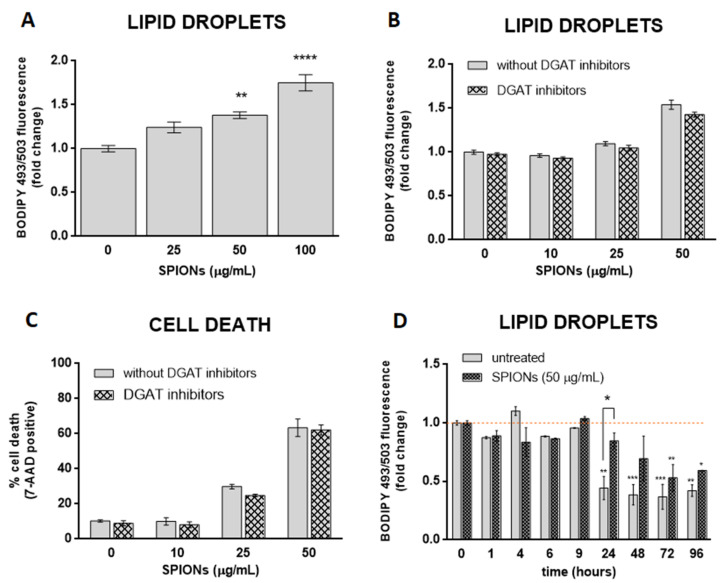
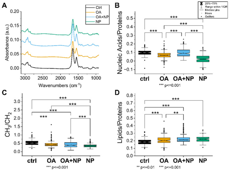
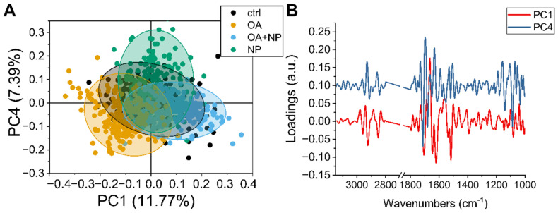
Superparamagnetic iron oxide nanoparticles (SPIONs) have great potential for use in medicine, but they may cause side effects due to oxidative stress. In our study, we investigated the effects of silica-coated SPIONs on endothelial cells and whether oleic acid (OA) can protect the cells from their harmful effects. We used viability assays, flow cytometry, infrared spectroscopy, fluorescence microscopy, and transmission electron microscopy. Our results show that silica-coated SPIONs are internalized by endothelial cells, where they increase the amount of reactive oxygen species (ROS) and cause cell death. Exposure to silica-coated SPIONs induced accumulation of lipid droplets (LD) that was not dependent on diacylglycerol acyltransferase (DGAT)-mediated LD biogenesis, suggesting that silica-coated SPIONs suppress LD degradation. Addition of exogenous OA promoted LD biogenesis and reduced SPION-dependent increases in oxidative stress and cell death. However, exogenous OA protected cells from SPION-induced cell damage even in the presence of DGAT inhibitors, implying that LDs are not required for the protective effect of exogenous OA. The molecular phenotype of the cells determined by Fourier transform infrared spectroscopy confirmed the destructive effect of silica-coated SPIONs and the ameliorative role of OA in the case of oxidative stress. Thus, exogenous OA protects endothelial cells from SPION-induced oxidative stress and cell death independent of its incorporation into triglycerides.

Keywords: endothelial cells; lipid droplets; oleic acid; oxidative stress; superparamagnetic iron oxide nanoparticles.


Conflict of interest statement

The authors declare no conflict of interest.

Figures

Figure 1
Figure 1
Particle characteristics and their internalization by endothelial cells. (A,B) Representative transmission electron microscopy (TEM) images of silica-coated superparamagnetic iron oxide nanoparticles (SPIONs) at low (A) and high magnification (B). (C) Zeta potential distribution of silica-coated SPIONs in serum-free medium. (D) Hydrodynamic size distribution of silica-coated SPIONs in serum-free medium. (E,F) The internalization of silica-coated SPIONs in human umbilical vein endothelial cells (HUVEC): representative TEM images of untreated HUVEC cells (E) and HUVEC cells exposed to silica-coated SPIONs at concentration 50 µg/mL for 24 h (F). Note numerous internalized electron-dense silica-coated SPIONs (red arrows) in autophagosomes.
Figure 2
Figure 2
Silica-coated SPIONs decrease cell viability and induce elevation of reactive oxygen species (ROS). (AC) HUVEC cells were treated with silica-coated SPIONs in serum-free medium for 24, 48, or 72 h. Cell viability was determined by resazurin assay (A), neutral red uptake (NRU) assay (B), and CyQuant assay (C). (D,E) HUVEC cells were treated with silica-coated SPIONs and N-acetylcysteine (NAC) (20 mM) or α-tocopherol (1 mM) for 24 h. Cell death (D) and ROS (E) were quantified by flow cytometry using 7-aminoactinomycin D (7-AAD) staining and CM-H2DCFDA staining, respectively. Values on the graphs are means ± standard error of the mean (SEM) of at least three independent experiments; statistically significant differences in mean values are indicated (*, p < 0.05; ***, p < 0.001; one-way ANOVA with Tukey post hoc test).
Figure 3
Figure 3
Silica-coated SPIONs-induced lipid droplet (LD) accumulation is not dependent on diacylglycerol acyltransferase (DGAT)-mediated LD biogenesis. (A) HUVEC cells were treated with silica-coated SPIONs (25, 50, and 100 µg/mL) for 24 h and LDs were quantified by flow cytometry using BODIPY 493/503 staining. (B,C) HUVEC cells were treated with combination of DGAT1 and DGAT2 inhibitors (5 µM T863 and 5 µM PF-06424439) and silica-coated SPIONs for 24 h. LDs and cell death were quantified by flow cytometry using BODIPY 493/503 (B) and 7-AAD staining (C), respectively. (D) HUVEC cells were treated with silica-coated SPIONs and LD levels were quantified in different time points (1–96 h) by flow cytometry using BODIPY 493/503 staining. Values on the graphs are means ± SEM of at least three independent experiments; statistically significant differences in mean values are indicated (*, p < 0.05; **, p < 0.01; ***, p < 0.001; ****, p < 0.0001; one-way ANOVA with Tukey post hoc test).
Figure 4
Figure 4
LD formation is not required for OA to inhibit silica-coated SPIONs-induced cell death and oxidative stress. (A) HUVEC cells were treated with 30, 50, and 100 µM OA for 24 h and LDs were quantified by flow cytometry, using BODIPY 493/503 staining. (B,C) Cells were treated with OA (100 µM) and silica-coated SPIONs (25 and 50 µg/mL) for 24 h. Cell death and ROS were quantified by flow cytometry using 7-AAD (B) and CM-H2DCFDA (C) staining, respectively. (D) Cells were treated with 100 µM OA and/or DGAT inhibitors (5 µM T863 and 5 µM PF-06424439) for 24 h. Cells were stained with BODIPY 493/503 and Hoechst 33342 to visualize LD (green) and nuclei (blue), respectively. (E,F) HUVEC cells were treated with DGAT inhibitors (5 µM T863 and 5 µM PF-06424439) and OA (30, 50, and 100 µM) for 24 h. LD and cell death were quantified by flow cytometry using BODIPY 493/503 (E) and 7-AAD (F) staining, respectively. (G) HUVEC cells were treated with DGAT inhibitors (5 µM T863 and 5 µM PF-06424439), OA (30 µM), and silica-coated SPIONs (50 µg/mL) for 24 h. Cell death was quantified by flow cytometry using 7-AAD staining. Values on the graphs are means ± SEM of at least three independent experiments; statistically significant differences in mean values are indicated (*, p < 0.05; **, p < 0.01; ***, p < 0.001; one-way ANOVA with Tukey post hoc test).
Figure 5
Figure 5
(A) Average spectra of the four analyzed cell groups after preprocessing, from bottom to top: Controls (CTRL) in black (average of 174 spectra), cells treated with oleic acid (OA) in yellow (average of 225 spectra), cells treated with OA+ silica-coated SPIONs (OA + nanoparticles (NP)) in blue (average of 280 spectra), and in green the average spectrum of SPION-treated cells (NP, average of 188 spectra). (B) Violin plot of the ratio of nucleic acid bands to total proteins. (C) Violin plot of the ratio of lipid bands to total amount of proteins. (D) Violin plot of the ratio of CH3 to CH2 bands of lipids. The same visualization parameters were used for all violin plots: CTRL in black, OA in yellow, OA + NP in blue, and NP in green. Values are sorted from the lowest value on the left to the highest on the right. Statistically significant differences in mean values are marked (**, p < 0.01; ***, p < 0.001).
Figure 6
Figure 6
(A) Scatter plot of principal component analysis (PCA) scores of principal component (PC)1 and PC4. (B) Line plot of charge vectors corresponding to PC1 (in black) and PC4 (in red). For clarity, an offset of 0.1 a.u. was added to the y-axis.

References

    1. Dulińska-Litewka J., Łazarczyk A., Hałubiec P., Szafrański O., Karnas K., Karewicz A. Superparamagnetic Iron Oxide Nanoparticles-Current and Prospective Medical Applications. Materials. 2019;12:617. doi: 10.3390/ma12040617. - DOI - PMC - PubMed
    1. Waddington D.E.J., Boele T., Maschmeyer R., Kuncic Z., Rosen M.S. High-Sensitivity in Vivo Contrast for Ultra-Low Field Magnetic Resonance Imaging Using Superparamagnetic Iron Oxide Nanoparticles. Sci. Adv. 2020;6:998–1015. doi: 10.1126/sciadv.abb0998. - DOI - PMC - PubMed
    1. Hedayatnasab Z., Dabbagh A., Abnisa F., Wan Daud W.M.A. Polycaprolactone-Coated Superparamagnetic Iron Oxide Nanoparticles for in Vitro Magnetic Hyperthermia Therapy of Cancer. Eur. Polym. J. 2020;133:109789. doi: 10.1016/j.eurpolymj.2020.109789. - DOI
    1. Samrot A.V., Sahithya C.S., Selvarani J., Purayil S.K., Ponnaiah P. A Review on Synthesis, Characterization and Potential Biological Applications of Superparamagnetic Iron Oxide Nanoparticles. Curr. Res. Green Sustain. Chem. 2021;4:100042. doi: 10.1016/j.crgsc.2020.100042. - DOI
    1. Patil R.M., Thorat N.D., Shete P.B., Bedge P.A., Gavde S., Joshi M.G., Tofail S.A.M., Bohara R.A. Comprehensive Cytotoxicity Studies of Superparamagnetic Iron Oxide Nanoparticles. Biochem. Biophys. Rep. 2018;13:63–72. doi: 10.1016/j.bbrep.2017.12.002. - DOI - PMC - PubMed