. 2022 Jun 25;23(13):7085.
doi: 10.3390/ijms23137085.

Regulation and Release of Vasoactive Endoglin by Brain Endothelium in Response to Hypoxia/Reoxygenation in Stroke

Affiliations

Regulation and Release of Vasoactive Endoglin by Brain Endothelium in Response to Hypoxia/Reoxygenation in Stroke

Axel Haarmann et al. Int J Mol Sci. .

Abstract

In large vessel occlusion stroke, recanalization to restore cerebral perfusion is essential but not necessarily sufficient for a favorable outcome. Paradoxically, in some patients, reperfusion carries the risk of increased tissue damage and cerebral hemorrhage. Experimental and clinical data suggest that endothelial cells, representing the interface for detrimental platelet and leukocyte responses, likely play a crucial role in the phenomenon referred to as ischemia/reperfusion (I/R)-injury, but the mechanisms are unknown. We aimed to determine the role of endoglin in cerebral I/R-injury; endoglin is a membrane-bound protein abundantly expressed by endothelial cells that has previously been shown to be involved in the maintenance of vascular homeostasis. We investigated the expression of membranous endoglin (using Western blotting and RT-PCR) and the generation of soluble endoglin (using an enzyme-linked immunosorbent assay of cell culture supernatants) after hypoxia and subsequent reoxygenation in human non-immortalized brain endothelial cells. To validate these in vitro data, we additionally examined endoglin expression in an intraluminal monofilament model of permanent and transient middle cerebral artery occlusion in mice. Subsequently, the effects of recombinant human soluble endoglin were assessed by label-free impedance-based measurement of endothelial monolayer integrity (using the xCELLigence DP system) and immunocytochemistry. Endoglin expression is highly inducible by hypoxia in human brain endothelial monolayers in vitro, and subsequent reoxygenation induced its shedding. These findings were corroborated in mice during MCAO; an upregulation of endoglin was displayed in the infarcted hemispheres under occlusion, whereas endoglin expression was significantly diminished after transient MCAO, which is indicative of shedding. Of note is the finding that soluble endoglin induced an inflammatory phenotype in endothelial monolayers. The treatment of HBMEC with endoglin resulted in a decrease in transendothelial resistance and the downregulation of VE-cadherin. Our data establish a novel mechanism in which hypoxia triggers the initial endothelial upregulation of endoglin and subsequent reoxygenation triggers its release as a vasoactive mediator that, when rinsed into adjacent vascular beds after recanalization, can contribute to cerebral reperfusion injury.

Keywords: CD105; HBMEC; endoglin; human brain endothelium; hypoxia; ischemia/reperfusion injury; middle cerebral artery occlusion; reoxygenation; soluble endoglin; stroke; vascular homeostasis.


Conflict of interest statement

The authors declare no conflict of interest.

Figures
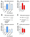
Figure 1
Figure 1
Hypoxia- and inflammation-modulated endoglin (ENG) expression in human brain endothelial cells (HBMEC). (A) ENG mRNA levels by quantitative real-time PCR in HBMEC after hypoxia (1% O2) or hypoxia/reoxygenation for the indicated durations relative to an unstimulated control. Data of six independent experiments and statistical analysis by the Kruskal–Wallis test followed by Dunn’s post-hoc test. (B) Representative Western blot on whole-cell extracts after stimulation as in A. (C) Quantification of ENG protein expression relative to β-actin in stimulated compared to unstimulated HBMEC of four independent experiments. Statistical testing for normal distribution by the D’Agostino–Pearson omnibus K2 test, followed by one-way ANOVA and the Bonferroni post-hoc test. (DF) ENG mRNA levels and ENG protein concentrations after stimulation with IL-1β (10 ng/mL) for 4 h and 24 h. Statistics as in B. Asterisks indicate significance compared to an unstimulated control (** p < 0.01; *** p < 0.001).
Figure 2
Figure 2
Increased matrix metalloproteinase-14 (MMP14) expression of human brain endothelium goes hand in hand with shedding of soluble endoglin (ENG). (A) MMP14 mRNA concentrations by quantitative real-time PCR in HBMEC after hypoxia (1% O2) or hypoxia/reoxygenation for the indicated durations relative to an unstimulated control. Data of three independent experiments. Statistical analysis by the Kruskal–Wallis test followed by Dunn’s post-hoc test. (B) Enzyme-linked immunosorbent assay for sENG in supernatants of HBMEC, stimulated as in A. Concentration relative to an untreated control. Statistical testing for normal distribution by the D’Agostino-Pearson omnibus K2 test, followed by one-way ANOVA and the Bonferroni post-test. (C) MMP14 mRNA concentrations after stimulation with IL-1β (10 ng/mL) for 4 h and 24 h; statistics as in A. (D) Concentration of sENG in cell culture supernatants after stimulation with IL-1β (10 ng/mL) for 4 h and 24 h; statistics as in B. (* p < 0.05; ** p < 0.01; *** p < 0.001).
Figure 3
Figure 3
The upregulation and release of ENG in permanent versus transient middle cerebral artery occlusion (MCAO) in a murine stroke model. Representative immunohistochemistry of cerebral ENG after 2 h (A) and 4 h (B) of permanent MCAO or after 2 h of MCAO with an additional 6 h of reperfusion (C). Sections show tissue 0.5 mm anterior from bregma. Scale bar = 1000 µm. (DF) Endothelial expression of ENG (red) on an individual vessel as identified by CD31 staining (green) of the basal ganglia region of the ischemic hemisphere in more detail. Scale bar = 50 µm. (G) Quantification of ENG expression in the basal ganglia depicted as the ratio of the mean fluorescence intensity between three corresponding regions of interest in ischemic vs. contralateral hemispheres, using ImageJ in n = 4 (2 h MCAO + 6 h reperfusion) and n = 5 (2 + 4 h MCAO) animals. (* p < 0.05; *** p < 0.001).
Figure 3
Figure 3
The upregulation and release of ENG in permanent versus transient middle cerebral artery occlusion (MCAO) in a murine stroke model. Representative immunohistochemistry of cerebral ENG after 2 h (A) and 4 h (B) of permanent MCAO or after 2 h of MCAO with an additional 6 h of reperfusion (C). Sections show tissue 0.5 mm anterior from bregma. Scale bar = 1000 µm. (DF) Endothelial expression of ENG (red) on an individual vessel as identified by CD31 staining (green) of the basal ganglia region of the ischemic hemisphere in more detail. Scale bar = 50 µm. (G) Quantification of ENG expression in the basal ganglia depicted as the ratio of the mean fluorescence intensity between three corresponding regions of interest in ischemic vs. contralateral hemispheres, using ImageJ in n = 4 (2 h MCAO + 6 h reperfusion) and n = 5 (2 + 4 h MCAO) animals. (* p < 0.05; *** p < 0.001).
Figure 4
Figure 4
Soluble ENG impairs resting endothelial monolayer integrity causing increased paracellular permeability and the downregulation of VE-cadherin. (A) Label-free assessment of transendothelial resistance of HBMEC in an impedance-based xCELLigence DP system. Confluent monolayers were stimulated with the indicated concentrations of sENG. Data represent the changes relative to untreated cells of five independent experiments. (B) Immunocytochemistry of resting HBMEC visualizing VE-cadherin after stimulation, as indicated. Representative of three experiments. Scale bar = 50 µm. (C) Quantification of VE-cadherin staining by blotting the ratio of the cell borders with positive staining to the cell circumference. Statistical testing for normal distribution by the D’Agostino–Pearson omnibus K2 test, followed by one-way ANOVA and the Bonferroni post-hoc test. (* p < 0.05; ** p < 0.01; *** p < 0.001).

References

    1. Mueller-Kronast N.H., Zaidat O.O., Froehler M.T., Jahan R., Aziz-Sultan M.A., Klucznik R.P., Saver J.L., Hellinger F.R., Jr., Yavagal D.R., Yao T.L., et al. Systematic Evaluation of Patients Treated With Neurothrombectomy Devices for Acute Ischemic Stroke: Primary Results of the STRATIS Registry. Stroke. 2017;48:2760–2768. doi: 10.1161/STROKEAHA.117.016456. - DOI - PubMed
    1. Knowland D., Arac A., Sekiguchi K.J., Hsu M., Lutz S.E., Perrino J., Steinberg G.K., Barres B.A., Nimmerjahn A., Agalliu D. Stepwise recruitment of transcellular and paracellular pathways underlies blood-brain barrier breakdown in stroke. Neuron. 2014;82:603–617. doi: 10.1016/j.neuron.2014.03.003. - DOI - PMC - PubMed
    1. Klohs J., Steinbrink J., Bourayou R., Mueller S., Cordell R., Licha K., Schirner M., Dirnagl U., Lindauer U., Wunder A. Near-infrared fluorescence imaging with fluorescently labeled albumin: A novel method for non-invasive optical imaging of blood-brain barrier impairment after focal cerebral ischemia in mice. J. Neurosci. Methods. 2009;180:126–132. doi: 10.1016/j.jneumeth.2009.03.002. - DOI - PubMed
    1. Jickling G.C., Liu D., Stamova B., Ander B.P., Zhan X., Lu A., Sharp F.R. Hemorrhagic transformation after ischemic stroke in animals and humans. J. Cereb. Blood Flow Metab. 2014;34:185–199. doi: 10.1038/jcbfm.2013.203. - DOI - PMC - PubMed
    1. Rossi E., Pericacho M., Bachelot-Loza C., Pidard D., Gaussem P., Poirault-Chassac S., Blanco F.J., Langa C., Gonzalez-Manchon C., Novoa J.M.L., et al. Human endoglin as a potential new partner involved in platelet-endothelium interactions. Cell. Mol. Life Sci. 2018;75:1269–1284. doi: 10.1007/s00018-017-2694-7. - DOI - PMC - PubMed