. 2022 Jun 25;23(13):7086.
doi: 10.3390/ijms23137086.

Cathepsin B p.Gly284Val Variant in Parkinson's Disease Pathogenesis

Affiliations

Cathepsin B p.Gly284Val Variant in Parkinson's Disease Pathogenesis

Lukasz M Milanowski et al. Int J Mol Sci. .

Abstract

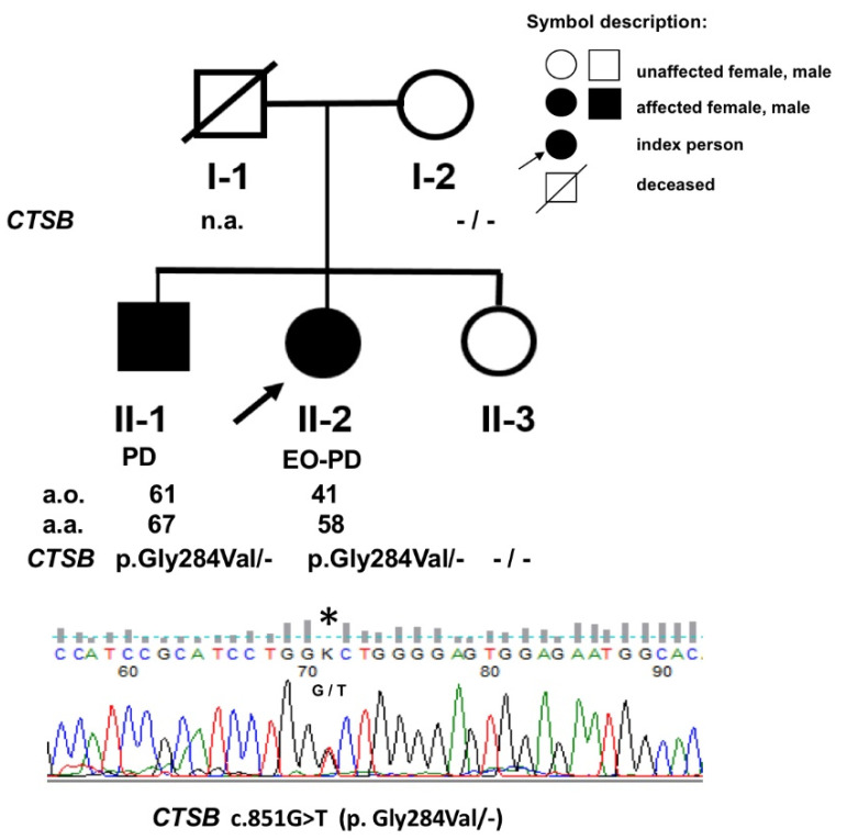
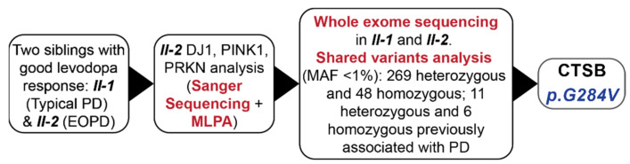
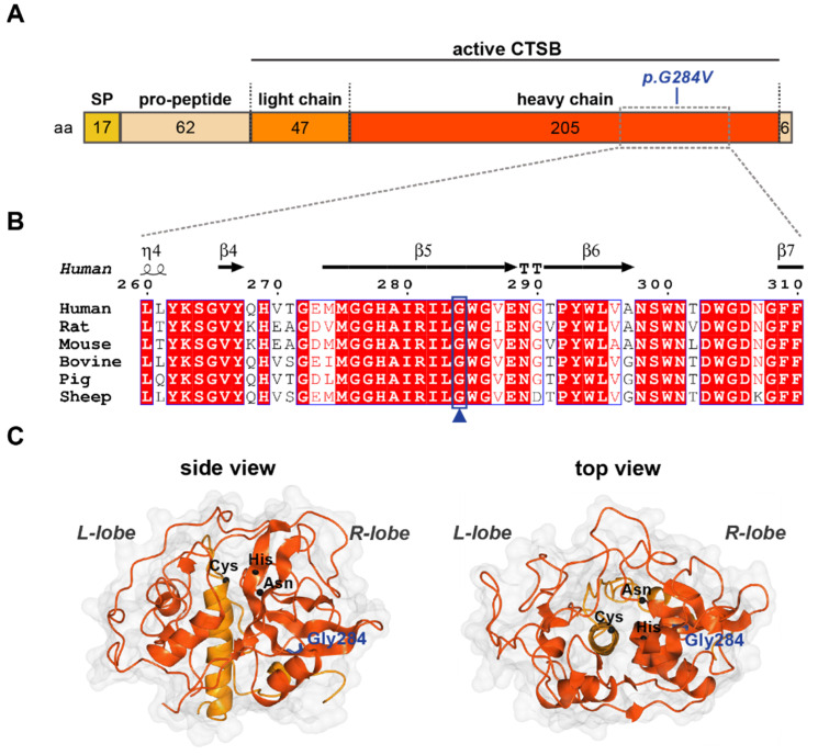
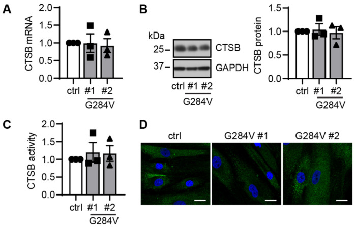
Parkinson's disease (PD) is generally considered a sporadic disorder, but a strong genetic background is often found. The aim of this study was to identify the underlying genetic cause of PD in two affected siblings and to subsequently assess the role of mutations in Cathepsin B (CTSB) in susceptibility to PD. A typical PD family was identified and whole-exome sequencing was performed in two affected siblings. Variants of interest were validated using Sanger sequencing. CTSB p.Gly284Val was genotyped in 2077 PD patients and 615 unrelated healthy controls from the Czech Republic, Ireland, Poland, Ukraine, and the USA. The gene burden analysis was conducted for the CTSB gene in an additional 769 PD probands from Mayo Clinic Florida familial PD cohort. CTSB expression and activity in patient-derived fibroblasts and controls were evaluated by qRT-PCR, western blot, immunocytochemistry, and enzymatic assay. The CTSB p.Gly284Val candidate variant was only identified in affected family members. Functional analysis of CTSB patient-derived fibroblasts under basal conditions did not reveal overt changes in endogenous expression, subcellular localization, or enzymatic activity in the heterozygous carrier of the CTSB variant. The identification of the CTSB p.Gly284Val may support the hypothesis that the CTSB locus harbors variants with differing penetrance that can determine the disease risk.

Keywords: CTSB; Parkinson’s disease; familial forms; fibroblasts; monogenic forms.


Conflict of interest statement

There is no conflict of interest relevant to the study.

Figures

Figure 1
Figure 1
Pedigree of the Polish PD family chosen for the study. (n.a.—not available, a.o.—age of disease onset, a.a.—age of patient’s analysis, *—the mutation site).
Figure 2
Figure 2
Flowchart of genetic experiments conducted on the typical PD family. EOPD = early onset Parkinson’s disease, MAF = minor allele frequency, MLPA—Multiplex ligation-dependent probe amplification.
Figure 3
Figure 3
Glycine 284 is conserved in CTSB across species. (A) Primary structure of CTSB indicating length of fragments in amino acids (aa) and position of the p.Gly284V mutation. SP—signal peptide. (B) Structure-informed multiple sequence alignment of six CTSB homologs. The secondary structure for human CTSB (PDB: 6AY2) is shown above. Boxed residues are conserved: white background with red text indicates functionally equivalent residues; red background with white text indicates sequence conservation. The blue box and arrowhead highlight Gly284, which is conserved across all species analyzed. (C) Crystal structure of mature human CTSB (PDB: 6AY2) shown in surface/ribbon representation, indicating the left (L) and right (R) lobes which constitute the mature enzyme. Heavy and light chains are colored red-orange and orange, respectively. Gly284 (blue) is shown in cylindrical representation; C-alphas of the catalytic triad: Cys (29) His (199) and Asn (219) are depicted as black spheres.
Figure 4
Figure 4
Functional analysis of CTSB in patient-derived skin cells. (A) qRT-PCR showed comparable mRNA levels in both CTSB mutant fibroblasts and the controls cells. (B) Representative western blot images of control and two CTSB mutant fibroblast lines (left). Western blot quantification showed similar levels of total CTSB protein levels in both CTSB mutant, and the WT control cells (right). (C) CTSB activity assay using cell lysates showed no differences in total enzymatic activity in the mutant fibroblast compared to the control. Circle—control, square—patient #1 carrying CTSB G284V, triangle—patient #2 carrying CTSB G284V. (D) Representative images of CTSB immunofluorescence staining (green) in fibroblasts at baseline condition. Scale bar: 20 µm. n = 3 independent experiments. Data are normalized to set control as 1 and are shown as mean with standard error. One-way ANOVA.

References

    1. Balestrino R., Schapira A. Parkinson disease. Eur. J. Neurol. 2020;27:27–42. doi: 10.1111/ene.14108. - DOI - PubMed
    1. Postuma R.B., Berg D., Stern M., Poewe W., Olanow C.W., Oertel W., Obeso J., Marek K., Litvan I., Lang A.E. MDS clinical diagnostic criteria for Parkinson’s disease. Mov. Disord. 2015;30:1591–1601. doi: 10.1002/mds.26424. - DOI - PubMed
    1. Dickson D.W. Neuropathology of Parkinson disease. Parkinsonism Relat. Disord. 2018;46:S30–S33. doi: 10.1016/j.parkreldis.2017.07.033. - DOI - PMC - PubMed
    1. Karimi-Moghadam A., Charsouei S., Bell B., Jabalameli M.R. Parkinson disease from mendelian forms to genetic susceptibility: New molecular insights into the neurodegeneration process. Cell. Mol. Neurobiol. 2018;38:1153–1178. doi: 10.1007/s10571-018-0587-4. - DOI - PMC - PubMed
    1. Nalls M.A., Blauwendraat C., Vallerga C.L., Heilbron K., Bandres-Ciga S., Chang D., Tan M., Kia D.A., Noyce A.J., Xue A. Identification of novel risk loci, causal insights, and heritable risk for Parkinson’s disease: A meta-analysis of genome-wide association studies. Lancet Neurol. 2019;18:1091–1102. doi: 10.1016/S1474-4422(19)30320-5. - DOI - PMC - PubMed