. 2022 Jun 27;23(13):7131.
doi: 10.3390/ijms23137131.

Cisplatin-Resistant CD44+ Lung Cancer Cells Are Sensitive to Auger Electrons

Affiliations

Cisplatin-Resistant CD44+ Lung Cancer Cells Are Sensitive to Auger Electrons

Karina Lindbøg Madsen et al. Int J Mol Sci. .

Abstract

Cancer stem cells (CSCs) are resistant to conventional therapy and present a major clinical challenge since they are responsible for the relapse of many cancers, including non-small cell lung cancer (NSCLC). Hence, future successful therapy should also eradicate CSCs. Auger electrons have demonstrated promising therapeutic potential and can induce DNA damage while sparing surrounding cells. Here, we sort primary patient-derived NSCLC cells based on their expression of the CSC-marker CD44 and investigate the effects of cisplatin and a thymidine analog (deoxyuridine) labeled with an Auger electron emitter (125I). We show that the CD44+ populations are more resistant to cisplatin than the CD44- populations. Interestingly, incubation with the thymidine analog 5-[125I]iodo-2'-deoxyuridine ([125I]I-UdR) induces equal DNA damage, G2/M cell cycle arrest, and apoptosis in the CD44- and CD44+ populations. Our results suggest that Auger electron emitters can also eradicate resistant lung cancer CD44+ populations.

Keywords: CD44; DNA damage; apoptosis; auger electrons; cancer stem cells; cisplatin; lung cancer.


Conflict of interest statement

The authors declare no conflict of interest. The funders had no role in the design of the study, in the collection, analyses, or interpretation of data, in the writing of the manuscript, or in the decision to publish the results.

Figures

Figure 1
Figure 1
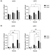
Tumorsphere formation and the relative expression of SOX2, NANOG, POU5F1, and CD44. (A) Generation of tumorspheres from a single cell. (B) Relative expression of pluripotency markers SOX2, NANOG, and POU5F1 and the surface marker CD44 in sorted LUC10 and LUC13 cells. Experiments were performed as three independent replicates. Values are expressed as the mean ± standard deviation. *: p < 0.05; ***: p < 0.001; and ****: p < 0.0001.
Figure 2
Figure 2
Doubling time and proliferation. (A) The doubling time for CD44-sorted cells grown as tumorspheres. (B) Upper panel: representative fluorescence microscopy images of CD44-sorted cells labeled with EdU (proliferation; green), PE-labeled anti-CD44 antibodies (red), and counterstained with DAPI (blue; magnification ×20). Lower panel: quantification of negative, EdU-positive, EdU- and CD44-positive, and CD44-positive cells. Experiments were performed as three independent replicates. Values are expressed as the mean ± standard deviation. *: p < 0.05 and ****: p < 0.0001.
Figure 3
Figure 3
Viability, DNA damage, and apoptosis (annexin V-positive cells) after cisplatin treatment. (A) The viability was evaluated using the CellTiter-Blue assay after 72 h incubation with increasing concentrations of cisplatin. (B) The percentage of DNA DSBs after treatment with 10 μM cisplatin for 2 h and (C) apoptosis after 72 h. Experiments were performed as three independent replicates. Values are expressed as the mean ± standard deviation. *: p < 0.05; **: p < 0.01; and ****: p < 0.0001.
Figure 4
Figure 4
Cellular uptake and DNA incorporation of [125I]I-UdR. (A) Cellular uptake of 18.5 kBq/mL [125I]I-UdR after 1, 4, and 7 h. (B) DNA incorporation of 18.5 kBq/mL [125I]I-UdR after 1, 4 and 7 h. Experiments were performed as three independent replicates. Values are expressed as the mean ± standard deviation. *: p < 0.05; **: p < 0.01; ***: p < 0.001; and ****: p < 0.0001.
Figure 5
Figure 5
Cell viability after [125I]I-UdR treatment. The viability was evaluated using the CellTiter-Blue assay after 7 days of incubation with increasing activity concentrations of [125I]I-UdR. Experiments were performed as three independent replicates. Values are expressed as the mean ± standard deviation. **: p < 0.01; ***: p < 0.001; and ****: p < 0.0001.
Figure 6
Figure 6
Radiation-induced DNA damage. The percentage of DNA DSBs after treatment with 2.5 kBq/mL [125I]I-UdR was evaluated by the analysis of phospho-H2AX after 7 days. Experiments were performed as three independent replicates. Values are expressed as the mean ± standard deviation. **: p < 0.01; ***: p < 0.001; and ****: p < 0.0001.
Figure 7
Figure 7
Radiation-induced changes in the cell cycle. Cell cycle analysis after treatment with 2.5 kBq/mL [125I]I-UdR for 7 days. Experiments were performed as three independent replicates. Values are expressed as the mean ± standard deviation.*: p < 0.05; **: p < 0.01; ***: p < 0.001; and ****: p < 0.0001.
Figure 8
Figure 8
Radiation-induced apoptosis (annexin V-positive cells). Apoptosis after treatment with 2.5 kBq/mL [125I]I-UdR evaluated by annexin V after 7 days. Experiments were performed as three independent replicates. Values are expressed as the mean ± standard deviation. **: p < 0.01; ****: p < 0.0001.
Figure 9
Figure 9
LUC10 and LUC13 were grown as tumorspheres. On the day of sorting, the tumorspheres were trypsinized and incubated with CD44 microbeads. The sorted cells were incubated with the Auger electron-emitting thymidine analog [125I]I-UdR for seven days, whereafter the viability, DNA damage, cell cycle, and apoptosis were analyzed.

References

    1. Sung H., Ferlay J., Siegel R.L., Laversanne M., Soerjomataram I., Jemal A., Bray F. Global Cancer Statistics 2020: GLOBOCAN Estimates of Incidence and Mortality Worldwide for 36 Cancers in 185 Countries. CA Cancer J. Clin. 2021;71:209–249. doi: 10.3322/caac.21660. - DOI - PubMed
    1. Travis W.D. Pathology of lung cancer. Clin. Chest Med. 2011;32:669–692. doi: 10.1016/j.ccm.2011.08.005. - DOI - PubMed
    1. Heng W.S., Gosens R., Kruyt F.A.E. Lung cancer stem cells: Origin, features, maintenance mechanisms and therapeutic targeting. Biochem. Pharmacol. 2019;160:121–133. doi: 10.1016/j.bcp.2018.12.010. - DOI - PubMed
    1. MacDonagh L., Gray S.G., Breen E., Cuffe S., Finn S.P., O’Byrne K.J., Barr M.P. Lung cancer stem cells: The root of resistance. Cancer Lett. 2016;372:147–156. doi: 10.1016/j.canlet.2016.01.012. - DOI - PubMed
    1. Leonetti A., Sharma S., Minari R., Perego P., Giovannetti E., Tiseo M. Resistance mechanisms to osimertinib in EGFR-mutated non-small cell lung cancer. Br. J. Cancer. 2019;121:725–737. doi: 10.1038/s41416-019-0573-8. - DOI - PMC - PubMed