. 2022 Jun 28;23(13):7200.
doi: 10.3390/ijms23137200.

Glioblastoma Extracellular Vesicle-Specific Peptides Inhibit EV-Induced Neuronal Cytotoxicity

Affiliations

Glioblastoma Extracellular Vesicle-Specific Peptides Inhibit EV-Induced Neuronal Cytotoxicity

Wenbo Zhou et al. Int J Mol Sci. .

Abstract

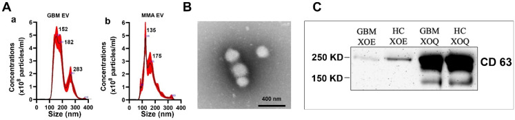
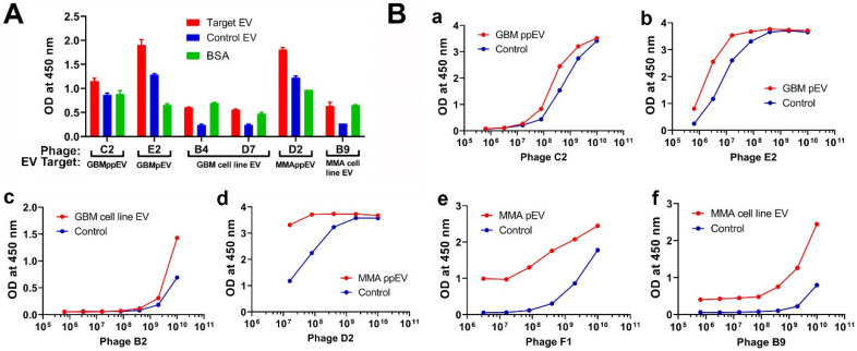
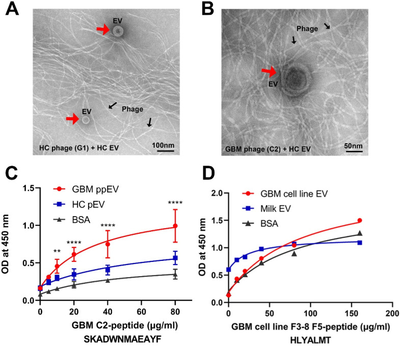
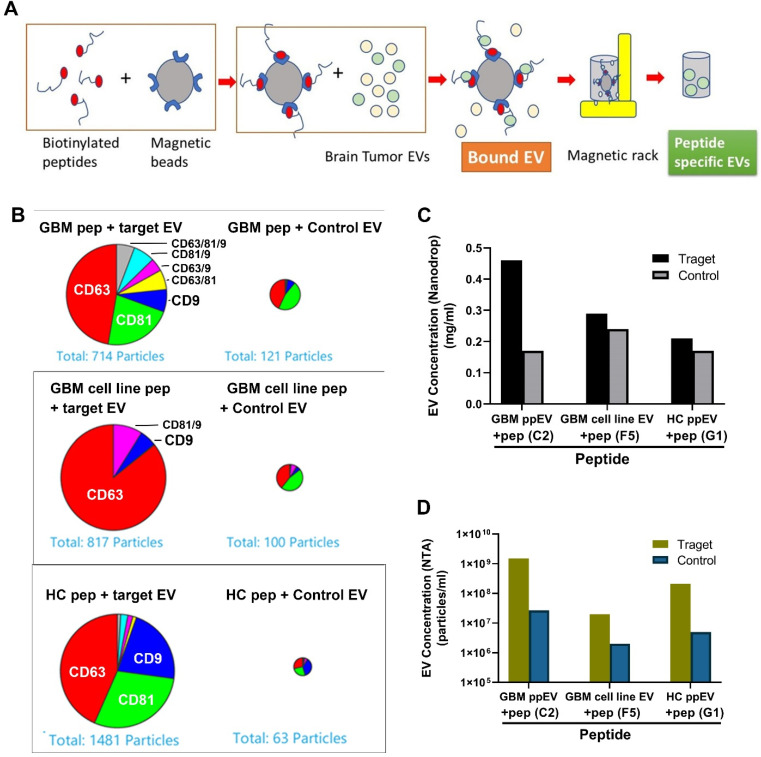
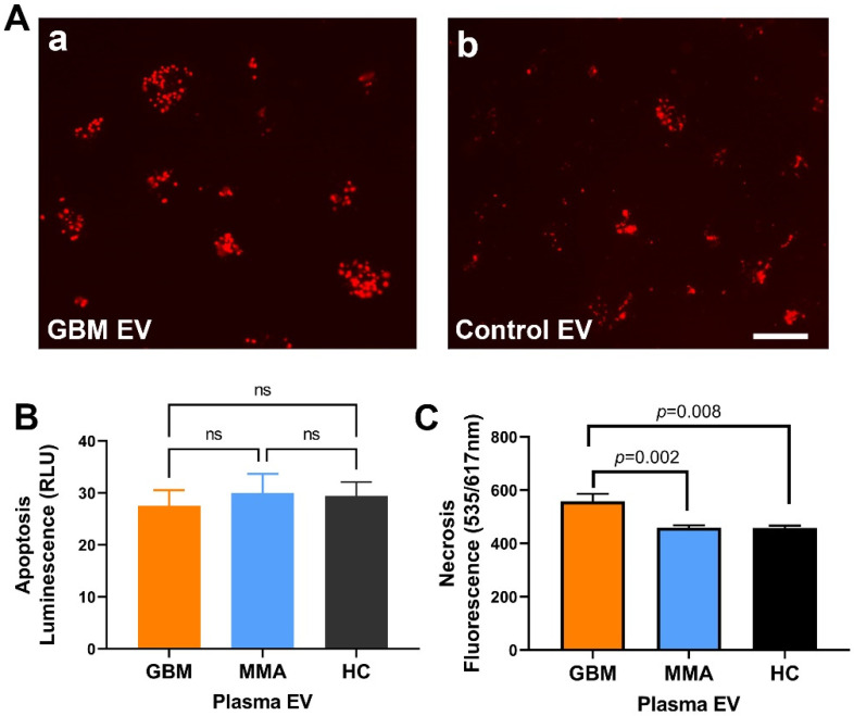
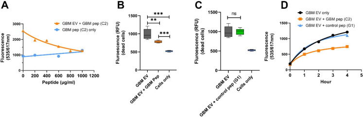
WHO Grade 4 IDH-wild type astrocytoma (GBM) is the deadliest brain tumor with a poor prognosis. Meningioma (MMA) is a more common "benign" central nervous system tumor but with significant recurrence rates. There is an urgent need for brain tumor biomarkers for early diagnosis and effective treatment options. Extracellular vesicles (EVs) are tiny membrane-enclosed vesicles that play essential functions in cell-to-cell communications among tumor cells. We aimed to identify epitopes of brain tumor EVs by phage peptide libraries. EVs from GBM plasma, MMA plasma, or brain tumor cell lines were used to screen phage-displayed random peptide libraries to identify high-affinity peptides. We purified EVs from three GBM plasma pools (23 patients), one MMA pool (10 patients), and four brain tumor cell lines. We identified a total of 21 high-affinity phage peptides (12 unique) specific to brain tumor EVs. The peptides shared high sequence homologies among those selected by the same EVs. Dose-response ELISA demonstrated that phage peptides were specific to brain tumor EVs compared to controls. Peptide affinity purification identified unique brain tumor EV subpopulations. Significantly, GBM EV peptides inhibit brain tumor EV-induced complement-dependent cytotoxicity (necrosis) in neurons. We conclude that phage display technology could identify specific peptides to isolate and characterize tumor EVs.

Keywords: ELISA; cytotoxicity; extracellular vesicles; glioblastoma; meningioma; neurons; peptides; phage-display; plasma; tumor cell line.


Conflict of interest statement

The authors declare no conflict of interest.

Figures

Figure 1
Figure 1
Characterization of EVs derived from plasma of patients with brain tumors. Plasma EVs were purified using ExoEasy and ExoQuick kits. (A) Nanoparticle Tracking Analysis showing examples of typical size and concentrations of the plasma EVs of GBM (a) and MMA (b) by ExoEasy. (B) Transmission electron microscope image showing ExoEasy purified GBM EVs, which are 100–200 nm diameters in size. (C) Western blots run on Native conditions demonstrated the presence of EV marker CD63 in GBM and HC plasma EVs. EVs were purified from plasma with ExoEasy (XOE) and ExoQuick (XOQ) followed by electrophoresis in Native gel. The blot was detected with mouse anti-human CD63 (1:1000). The CD63 proteins showed a much large size at 250 KD due to heavy glycosylation under native conditions, rather than the 25 KD under denaturing conditions. We found that CD63 protein was more abundant in Exo-Quick-purified EVs than in Exo-Easy-purified EVs.
Figure 2
Figure 2
ELISA demonstrates that phage peptides selected by brain tumor extracellular vesicles are specific to panning EVs. (A) Direct ELISA shows that phage peptides are specific to panning brain tumor EVs. The target EVs include GBM and MMA ppEVs (from pooled plasma), pEVs (from individual plasma), and tumor cell line EVs. EVs coated on a high-binding ELISA plate (50 mg/mL) were incubated with corresponding phage (2 × 10⁹ pfu/well). Bound phage peptides were detected by mouse anti-M13 pIII-HRP antibody (1:5000 dilution) followed by TMB substrate color detection. Each representative phage peptide (labeled in letters) was shown for OD values to corresponding panning EVs (target EV), control EVs, and BCA. (B) Dose–response ELISA demonstrating specific bindings of phage peptides to panning brain tumor EVs. GBM (B-ac) and MMA EVs (B-df) (pooled plasma, individual plasma, and tumor cell line EVs) at 50 mg/mL were directly coated onto wells of ELISA plates, followed by incubation with corresponding phage peptides in serial 5-fold dilutions starting with 1 × 10¹⁰ pfu/well. Detection was the same as described above. HC plasma EVs were used as controls for brain tumor plasma EVs (a,b,d,e), and normal human astrocyte EVs were used as controls for brain tumor EVs (c,f).
Figure 3
Figure 3
High-affinity bindings of phage peptides to GBM EVs are demonstrated by electron microscopy and synthetic peptide ELISA. (A,B) Electron microscopic images showing EVs precipitated by phage peptides. GBM EV phage C2 and HC EV phage G1 (2 × 10¹⁰ pfu) were incubated with HC-EVs (100 µg) overnight at 4 °C. The EV–phage complexes were precipitated by PEG 8000/NaCl, dissolved in PBS followed by transmission electron microscopy. (A) HC G1 phage and HC EVs. (B) GBM C2 phage and HC EVs. The EVs (red arrows) precipitated by M13 phage (black arrows) show typical spherical shapes with size ranges of 50–200 nm in diameter. Notice that samples in image (A) are 1:20 dilution and samples in (B) are original with no dilution, indicating that there are fewer HC EVs precipitated by GBM phage C2 compared to HC EVs by HC phage. (C,D) ELISA shows dose responses of synthetic peptide binding to corresponding EVs. EVs (20 µg/mL) and BSA protein (20 µg/mL) were coated onto wells of ELISA plates overnight at 4 °C. Synthetic biotinylated peptides in 2-fold serial dilutions were added to corresponding wells. Bound peptides were detected by NeutrAvidin HRP (1:10,000) followed by the TMB color reaction. (C) GBM plasma EV C2 peptide had significantly higher bindings to corresponding GBM ppEV compared to HC-EV or BSA (** p< 0.01, **** p < 0.0001). (D) GBM tumor cell line EV peptide demonstrated higher bindings to corresponding GBM cell line EVs compared to bovine milk EVs or BSA.
Figure 4
Figure 4
A higher number of GBM EVs was precipitated by EV-specific synthetic peptides. (A) Schematic drawing of EV precipitation with peptide magnetic beads. The biotinylated peptides bound to streptavidin linked Dynabeads were incubated with EVs at 4 °C overnight, and EVs were eluted with low pH buffer and neutralized with 1 M Tris-HCl (pH 10). (B) ExoView analysis demonstrated that higher amounts of EVs were affinity-precipitated by corresponding peptides compared to control peptides. GBM plasma EVs (top panel), GBM tumor cell line EVs (middle panel), and HC plasma EVs (lower panel) were precipitated by corresponding peptides and control peptides, followed by analysis with ExoView. About 5- to 20-fold more target EVs were precipitated by peptides compared to control EVs. (C) Higher target EV concentrations precipitated by specific peptides compared to control EVs. (D) NTA showed that higher target EV concentrations were precipitated by specific peptides compared to control EVs. Specific peptides can capture 10–50 times more target EVs than control EVs.
Figure 5
Figure 5
GBM EV induced necrotic cell death in neurons (A). Fluorescent microscopy shows higher neuronal death induced by GBM EVs. SH-SY5Y cells were treated with EVs for one hour. Cell death was monitored by propidium iodide. A higher number of neuron death was seen in GBM EVs (A-a) as compared to control EVs from an epithelial cell line (A-b). (B). GBM EV did not cause apoptosis in SH-SY5Y cells. SH-SY5Y cells were treated with GBM, MMA, or HC EVs for four hours, and apoptosis was measured by luminescence. Only minimal luminescence signals were detected in GBM, MMA or HC EV-treated cells, and there are no significant differences among different EVs (n.s., p > 0.1, n = 6 for GBM EV, n = 8 for MMA EV, n = 5 for HC EV). (C). GBM plasma EVs induced higher necrosis as compared to control EVs. SH-SY5Y cells were incubated with plasma EVs (2 mg/mL, GBM EV=14; MMA EV = 8; HC EV = 8) for 4 h. The necrotic cell death was monitored by PI red fluorescent reading. There were significantly higher levels of necrosis in GBM-EV-treated neurons compared to MMA EV and HC-EV-treated cells (p = 0.002 and p = 0.008, respectively). Scale bar = 200 mm for (A).
Figure 6
Figure 6
GBM EV-specific peptides inhibit neuronal cytotoxicity induced by the GBM EVs. (A). GBM EV-specific peptide inhibits EV-induced neuronal cytotoxicity in a dose-dependent manner. SH-SY5Y cells were incubated with GBM pooled plasma EVs at 20 mg/mL plus 5% Normal Human Serum. GBM EV-specific peptide C2 in various amounts was added to cells at the same time as the EV treatment. Cell death was monitored by ethidium homodimer-1 (red fluorescent reading). With 1000 mg/mL peptide, the GBM EV peptide nearly completely blocked the GBM-EV cytotoxicity after 4-h treatment. Cells with peptides (C2) alone did not produce cytotoxicity. (B,C). GBM EV-specific peptides (B), but not the control peptides (C) show time-dependent inhibition of EV induced neuronal death. Neuroblastoma SH-SY5Y cells were treated with GBM EVs plus peptides, and cytotoxicity was monitored for 4 h. GBM EV-specific peptide C2 inhibited GBM-EV induced cytotoxicity (GBM EV + GBM Pep), but not the control peptide to HC EV (G1) (GBM EV + control pep G1) (** p < 0.01, *** p < 0.001). (D) Time course of GBM-C2 peptide for the inhibition of GBM-EV cytotoxicity. SH-SY5Y cells were treated with GBM EVs at 20 mg/mL plus 5% Normal Human Serum. The GBM-C2 peptide (1000 mg/mL) or the HC-G1 peptide (as control, 1000 mg/mL) was added to cells during the same time as EV treatment. The cell death was monitored by propidium iodide red fluorescent reading once per hour. Results showed that the GBM peptide can inhibit GBM EV-induced cell death.

References

    1. Louis D.N., Perry A., Wesseling P., Brat D.J., Cree I.A., Figarella-Branger D., Hawkins C., Ng H.K., Pfister S.M., Reifenberger G., et al. The 2021 WHO Classification of Tumors of the Central Nervous System: A summary. Neuro-Oncology. 2021;23:1231–1251. doi: 10.1093/neuonc/noab106. - DOI - PMC - PubMed
    1. Alexander B.M., Cloughesy T.F. Adult Glioblastoma. J. Clin. Oncol. 2017;35:2402–2409. doi: 10.1200/JCO.2017.73.0119. - DOI - PubMed
    1. Poon M.T.C., Sudlow C.L.M., Figueroa J.D., Brennan P.M. Longer-term (≥2 years) survival in patients with glioblastoma in population-based studies pre- and post-2005: A systematic review and meta-analysis. Sci. Rep. 2020;10:1–10. doi: 10.1038/s41598-020-68011-4. - DOI - PMC - PubMed
    1. Huntoon K., Toland A.M.S., Dahiya S. Meningioma: A Review of Clinicopathological and Molecular Aspects. Front. Oncol. 2020;10:579599. doi: 10.3389/fonc.2020.579599. - DOI - PMC - PubMed
    1. Preusser M., Brastianos P., Mawrin C. Advances in meningioma genetics: Novel therapeutic opportunities. Nat. Rev. Neurol. 2018;14:106–115. doi: 10.1038/nrneurol.2017.168. - DOI - PubMed