. 2022 Jun 29;23(13):7252.
doi: 10.3390/ijms23137252.

Extracellular Cysteines Are Critical to Form Functional Cx46 Hemichannels

Affiliations

Extracellular Cysteines Are Critical to Form Functional Cx46 Hemichannels

Ainoa Fernández-Olivares et al. Int J Mol Sci. .

Abstract

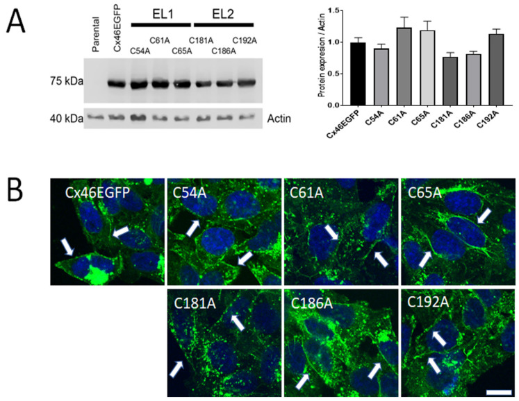
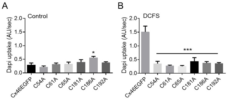
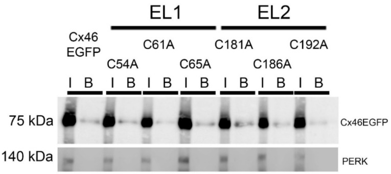
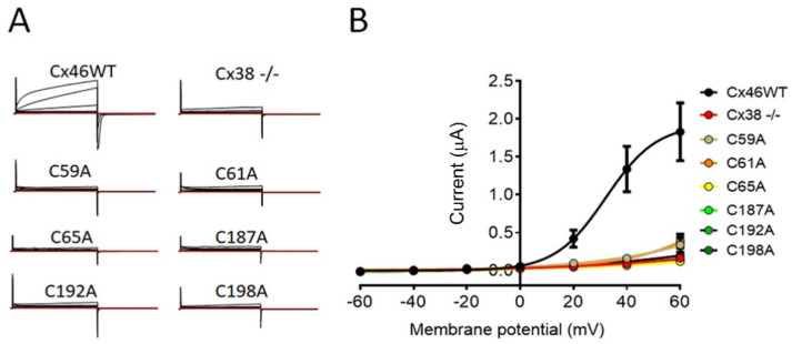
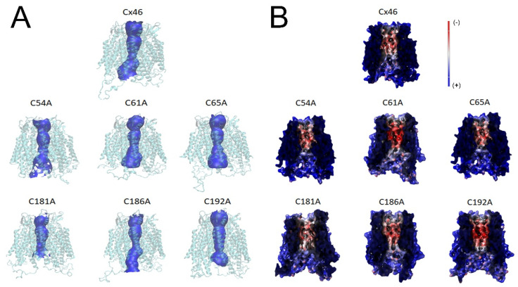
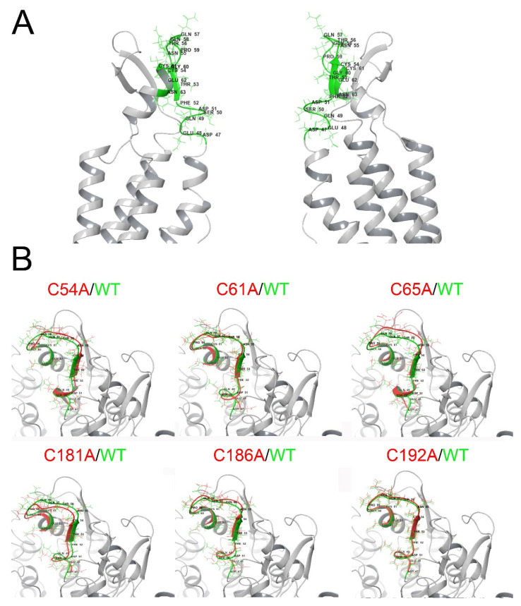
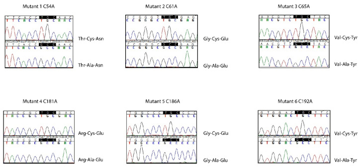
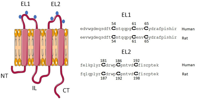
Connexin (Cxs) hemichannels participate in several physiological and pathological processes, but the molecular mechanisms that control their gating remain elusive. We aimed at determining the role of extracellular cysteines (Cys) in the gating and function of Cx46 hemichannels. We studied Cx46 and mutated all of its extracellular Cys to alanine (Ala) (one at a time) and studied the effects of the Cys mutations on Cx46 expression, localization, and hemichannel activity. Wild-type Cx46 and Cys mutants were expressed at comparable levels, with similar cellular localization. However, functional experiments showed that hemichannels formed by the Cys mutants did not open either in response to membrane depolarization or removal of extracellular divalent cations. Molecular-dynamics simulations showed that Cys mutants may show a possible alteration in the electrostatic potential of the hemichannel pore and an altered disposition of important residues that could contribute to the selectivity and voltage dependency in the hemichannels. Replacement of extracellular Cys resulted in "permanently closed hemichannels", which is congruent with the inhibition of the Cx46 hemichannel by lipid peroxides, through the oxidation of extracellular Cys. These results point to the modification of extracellular Cys as potential targets for the treatment of Cx46-hemichannel associated pathologies, such as cataracts and cancer, and may shed light into the gating mechanisms of other Cx hemichannels.

Keywords: channel permeability; connexins; extracellular loops; post-translational modification; redox sensing.


Conflict of interest statement

The authors declare no conflict of interest.

Figures

Figure 1
Figure 1
Expression and cellular localization of Cx46 Cys mutants. HeLa cells were transfected with a wild-type or Cys–Ala-mutated human Cx46 DNA fused to EGFP. (A) Representative blot of three independent Western blots. Fifty μg of protein were loaded in each line. A ~75-kDa band was detected in all the cases, except for non-transfected HeLa cells (Parental). Actin was used as loading control. The graph on the right presents the densitometric analysis of the three Western blots and shows no statistical differences between protein levels, when comparing Cys mutants and wild-type Cx46 (n = 3, n.s = p > 0.05). (B) Representative images of EGFP fluorescence in HeLa cells transfected with wild-type Cx46 or Cys mutants; nuclei were visualized with DAPI. White arrow heads denote fluorescence at cell-to-cell contacts. Calibration bar = 10 µm.
Figure 2
Figure 2
Hemichannels formed by Cys mutants do not open upon exposure to divalent cation free solution (DCFS). HeLa cells transfected with wild-type Cx46 or Cys mutants were placed in recording media containing 10 μM DAPI under control conditions or in DCFS. Pictures of 16 cells were taken every 20 s for 20 min, with 4 replicates. DAPI uptake was calculated as arbitrary units per second (AU/Sec). (A) Control conditions. The rate of DAPI uptake was similar in cells expressing all Cys mutants, except for the C186A mutant, which showed a small, but significant higher, DAPI uptake compared to cells expressing wild-type Cx46 (*, p < 0.05). (B) DCFS. HeLa cells expressing wild-type Cx46 showed an enhanced rate of DAPI uptake, which was not observed in any of the Cys mutants (***, p < 0.001) (n = 4 for each condition).
Figure 3
Figure 3
Cys mutants are present at the plasma membrane. Cell-surface biotinylation was performed in cultures of HeLa cells transfected with wild-type Cx46 or Cys mutants, and samples were then analyzed by Western blotting. I (Input): 50 μg of protein before the separation with streptavidin-magnetic beads; B: the whole sample after the separation with streptavidin-magnetic beads. The presence of the cytoplasmatic protein PERK was used as negative control and, as expected, was found only before separation and not in the biotinylated sample. (n = 3 independent experiments).
Figure 4
Figure 4
Hemichannels formed by Cys mutants are insensitive to membrane depolarization. Xenopus oocytes were injected with an oligo against the endogenous Cx38 alone (Cx38 -/-) or in combination with cRNA of wild-type Cx46 or Cys mutants. Hemichannel currents were recorded using dual-electrode voltage clamp after 24 h in 8 oocytes. (A) Representative recordings showing that only hemichannels formed of wild-type Cx46 open after membrane depolarization. (B) I/V plot summarizing results of the 8 independent experiments. The Boltzmann equation was used to fit current values.
Figure 5
Figure 5
MD simulations suggest discreet changes in both pore size and electrostatic potential in hemichannels formed by Cys mutants. Human Cx46 50 ns MD models. (A) Hemichannel tertiary structure is shown in light blue, while the pore profile is shown in blue. (B) Electrostatic potential of the hemichannel pore with negative and positive potentials represented in red and blue colors, respectively. Note that small red changes are observed in all the mutants, which were more evident in C61A and C192A.
Figure 6
Figure 6
MD simulations suggest changes in the 3D disposition of the TM1-EL1 parahelix segment. Human Cx46 50-ns MD models. (A) Representation of a Cx46 hemichannel showing the location of the parahelix (green segment). (B) Zoom of the parahelix segment (the segment between amino acids 47 and 61 was analyzed). The configuration of this segment in the wild-type Cx46 (green) is superimposed on the parahelix model for each Cys mutant (red).
Figure 7
Figure 7
Sequencing results from Cx46 Cys mutants. cDNA plasmids containing the mutagenesis products were sequenced. For each panel, the upper electropherogram shows the sequence for human wild-type Cx46, and the bottom part shows the sequence obtained for the mutants.
Figure 8
Figure 8
Schematic representation of the localization of extracellular Cys of Cx46. The diagram shows the topological disposition of Cx46 at the plasma membrane and the approximate location of extracellular Cys residues (blue dots). In this study, we used Cx46 derived from human and rat, where Cys located at the EL1 are identical, however, the Cys at EL2 show differences in location within the primary sequence, which are homologous in function.

References

    1. Sáez J.C., Retamal M., Basilio D., Bukauskas F.F., Bennett M.V. Connexin-based gap junction hemichannels: Gating mechanisms. Biochim. Biophys. Acta (BBA)-Biomembr. 2005;1711:215–224. doi: 10.1016/j.bbamem.2005.01.014. - DOI - PMC - PubMed
    1. Gong X.-Q., Nicholson B.J. Size Selectivity between Gap Junction Channels Composed of Different Connexins. Cell Commun. Adhes. 2001;8:187–192. doi: 10.3109/15419060109080721. - DOI - PubMed
    1. Myers J., Haddad B.G., O’Neill S.E., Chorev D., Yoshioka C.C., Robinson C., Zuckerman D.M., Reichow S.L. Structure of native lens connexin 46/50 intercellular channels by cryo-EM. Nature. 2018;564:372–377. doi: 10.1038/s41586-018-0786-7. - DOI - PMC - PubMed
    1. Maeda S., Nakagawa S., Suga M., Yamashita E., Oshima A., Fujiyoshi Y., Tsukihara T. Structure of the connexin 26 gap junction channel at 3.5 Å resolution. Nature. 2009;458:597–602. doi: 10.1038/nature07869. - DOI - PubMed
    1. Sáez J.C., Schalper K.A., Retamal M., Orellana J.A., Shoji K.F., Bennett M.V. Cell membrane permeabilization via connexin hemichannels in living and dying cells. Exp. Cell Res. 2010;316:2377–2389. doi: 10.1016/j.yexcr.2010.05.026. - DOI - PubMed

LinkOut - more resources