Review
. 2022 Jul 2;23(13):7377.
doi: 10.3390/ijms23137377.

Ca2+ Signalling and Hypoxia/Acidic Tumour Microenvironment Interplay in Tumour Progression

Affiliations
Review

Ca2+ Signalling and Hypoxia/Acidic Tumour Microenvironment Interplay in Tumour Progression

Madelaine Magalì Audero et al. Int J Mol Sci. .

Abstract

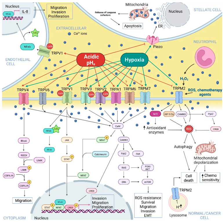
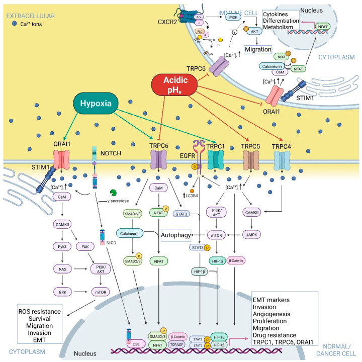
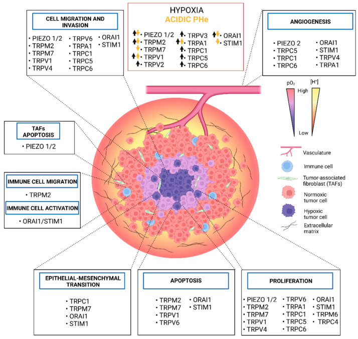
Solid tumours are characterised by an altered microenvironment (TME) from the physicochemical point of view, displaying a highly hypoxic and acidic interstitial fluid. Hypoxia results from uncontrolled proliferation, aberrant vascularization and altered cancer cell metabolism. Tumour cellular apparatus adapts to hypoxia by altering its metabolism and behaviour, increasing its migratory and metastatic abilities by the acquisition of a mesenchymal phenotype and selection of aggressive tumour cell clones. Extracellular acidosis is considered a cancer hallmark, acting as a driver of cancer aggressiveness by promoting tumour metastasis and chemoresistance via the selection of more aggressive cell phenotypes, although the underlying mechanism is still not clear. In this context, Ca2+ channels represent good target candidates due to their ability to integrate signals from the TME. Ca2+ channels are pH and hypoxia sensors and alterations in Ca2+ homeostasis in cancer progression and vascularization have been extensively reported. In the present review, we present an up-to-date and critical view on Ca2+ permeable ion channels, with a major focus on TRPs, SOCs and PIEZO channels, which are modulated by tumour hypoxia and acidosis, as well as the consequent role of the altered Ca2+ signals on cancer progression hallmarks. We believe that a deeper comprehension of the Ca2+ signalling and acidic pH/hypoxia interplay will break new ground for the discovery of alternative and attractive therapeutic targets.

Keywords: Ca2+ signalling; PIEZO channels; SOC channels; TRP channels; hypoxia; tumour acidic microenvironment; tumour progression.


Conflict of interest statement

The authors declare no conflict of interest.

Figures

Figure 1
Figure 1
Overview of the effects of acidic pHe and hypoxia on TRP and Piezo Ca2+-permeable channels. The positive or negative effects of hypoxia and acidic pHe on the Ca2+-permeable channels were obtained from both normal and cancer cells-related studies, while the signalling pathways indicated were obtained uniquely from cancer cells-based investigations. The figure depicts Piezo-, TRPV-, TRPA1- and TRPM-mediated Ca2+-dependent signalling pathways activated or inhibited by acidic pHe and hypoxia and linked to tumour progression. TRPV (TRPV1–4, 6), TRPA1, and TRPM (TRPM6, 7) expressed in cell cancer’s plasma membrane are differentially regulated by acidic pHe, being mostly activated by tumour acidosis, and transducing its signals to activate Ca2+-dependent downstream effectors, such as NF-κB, JAK/STAT, PI3K/AKT, NFAT, ERK, and LIMK. TRPA1 is also activated by hypoxia. These effectors promote tumour cell migration, invasion, proliferation, survival, mesenchymal phenotype, and chemoresistance. TRPV6 channels’ activity is inhibited by tumour acidosis, as TRPM2, which inhibition avoids induction of cancer cell death and reduces chemosensitivity. Piezo channels embedded in stellate cells’ plasma membrane are inhibited by acidic pHe, promoting stellate cells’ survival. TRPV1 activation in lymphatic endothelial cells promotes activation of NF-κB and upregulation of IL-8, a lymphangiogenic factor. CaM, calmodulin; CAMKII, Ca2+/calmodulin-dependent protein kinase II; Pyk2, protein tyrosine kinase 2; RAS, Rat sarcoma virus; ERK, extracellular signal-regulated kinase; FAK, Focal Adhesion Kinase; PI3K, phosphoinositide 3-kinase; AKT, protein kinase B; mTOR, mammalian target of rapamycin; NF-κB, nuclear factor-κB; JAK, Janus kinases; STAT, signal transducer and activator of transcription; NFAT, nuclear factor of activated T-cells; RhoA, Ras homolog family member A; ROCK, Rho-associated protein kinase; LIMK, LIM domain kinase; CREB, C-AMP response element-binding protein. The question mark indicates contradictory results in the literature. Created with BioRender.com, accessed on 20 June 2022.
Figure 2
Figure 2
Overview of the effects of acidic pHe and hypoxia on TRPC and ORAI Ca2+-permeable channels. The positive or negative effects of hypoxia and acidic pHe on the Ca2+-permeable channels were obtained from both normal and cancer cell-related studies, while the signalling pathways indicated were obtained uniquely from cancer cell-based investigations. The figure depicts TRPCs- and SOCs-mediated Ca2+-dependent signalling pathways inhibited or activated by acidic pHe or hypoxia and linked to tumour progression. TRPC (TRPC1, 4, 5) expressed in cancer cells’ plasma membrane is all activated by acidic pHe or hypoxia, transducing their signals to activate Ca2+-dependent downstream effectors, such as SMAD2/3, NFAT, STAT3, HIF1, AMPK and β-catenin. These effectors promote tumour cell migration, angiogenesis, invasion, proliferation, mesenchymal phenotype and chemoresistance and the expression of TRPC1, via EGFR activation, and TRPC6 via Notch1 signalling pathway, in a mechanism of positive feedback regulation for both TRPC1 and TRPC6 channels. Immune cells expressing TRPC6 channels on plasma membrane show TRPC6’s activity that is inhibited by acidic pHe, reducing their migration. ORAI1 channels function in immune cells is also negatively affected by acidic pHe, impairing different processes needed for immune cells’ anti-tumour activity. Hypoxia promotes both ORAI1 expression, via Notch signalling pathway, and activation, leading to increased ROS resistance, migration, invasion, EMT and cell survival. CaM, calmodulin; CAMKII, Ca2+/calmodulin-dependent protein kinase II; Pyk2, protein tyrosine kinase 2; RAS, rat sarcoma virus; ERK, extracellular signal-regulated kinase; FAK, focal adhesion kinase; PI3K, phosphoinositide 3-kinase; AKT, protein kinase B; mTOR, mammalian target of rapamycin; NICD, Notch intracellular domain; CSL, CBF1, suppressor of hairless, Lag-1; NFAT, nuclear factor of activated T-cells; STAT, signal transducers and activators of transcription; EGFR, epidermal growth factor receptor; HIF-1, hypoxia-inducible factor 1. Created with BioRender.com, accessed on 20 June 2022.
Figure 3
Figure 3
Schematic illustration of tumour microenvironment landscape. The increased tumour growth, the acidification of the extracellular space and aberrant vascularisation and limited O2 supply origin a tumour core that is hypoxic and acidic, with limited supply of oxygen and nutrients from the blood vessels. Peripheral tumour cells are located in regions with a higher extracellular pH, a result of proximity to blood vessels and the possibility to wash out acidic waste products. TRP, Piezo and SOCs channels expressed in cancer, immune and stromal cells are presented in the corresponding black boxes with indication of their involvement in different cancer hallmarks, such as proliferation, migration, invasion, angiogenesis, and epithelial–mesenchymal transition. The red box contains the up-to-date information regarding the effect (up arrows = positive effect; down arrows = negative effect) of hypoxia and acid pHe on the activity and/or expression of calcium-permeable channels in cancer cells or tumour-associated cells.

References

    1. Wei R., Liu S., Zhang S., Min L., Zhu S. Cellular and Extracellular Components in Tumour Microenvironment and Their Application in Early Diagnosis of Cancers. Anal. Cell. Pathol. 2020;2020:6283796. doi: 10.1155/2020/6283796. - DOI - PMC - PubMed
    1. Warburg O. On the Origin of Cancer Cells. Science. 1956;123:309–314. doi: 10.1126/science.123.3191.309. - DOI - PubMed
    1. Cassim S., Vučetić M., Ždralević M., Pouyssegur J. Warburg and Beyond: The Power of Mitochondrial Metabolism to Collaborate or Replace Fermentative Glycolysis in Cancer. Cancers. 2020;12:1119. doi: 10.3390/cancers12051119. - DOI - PMC - PubMed
    1. Marchiq I., Le Floch R., Roux D., Simon M.-P., Pouyssegur J. Genetic Disruption of Lactate/H + Symporters (MCTs) and Their Subunit CD147/BASIGIN Sensitizes Glycolytic Tumour Cells to Phenformin. Cancer Res. 2015;75:171–180. doi: 10.1158/0008-5472.CAN-14-2260. - DOI - PubMed
    1. Ždralević M., Brand A., Di Ianni L., Dettmer K., Reinders J., Singer K., Peter K., Schnell A., Bruss C., Decking S.-M., et al. Double Genetic Disruption of Lactate Dehydrogenases A and B Is Required to Ablate the “Warburg Effect” Restricting Tumour Growth to Oxidative Metabolism. J. Biol. Chem. 2018;293:15947–15961. doi: 10.1074/jbc.RA118.004180. - DOI - PMC - PubMed