. 2022 Jul 3;23(13):7408.
doi: 10.3390/ijms23137408.

Do Extracellular Vesicles Derived from Mesenchymal Stem Cells Contain Functional Mitochondria?

Affiliations

Do Extracellular Vesicles Derived from Mesenchymal Stem Cells Contain Functional Mitochondria?

Ljubava D Zorova et al. Int J Mol Sci. .

Abstract

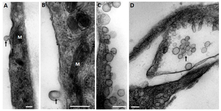
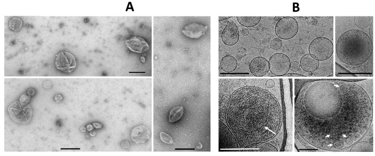
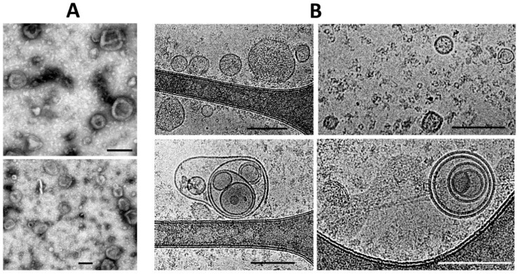
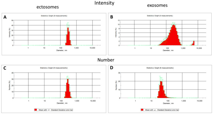
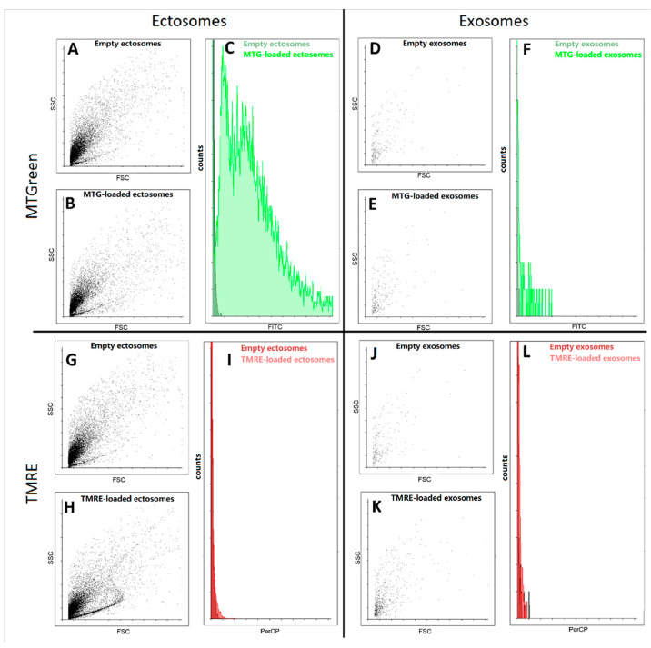
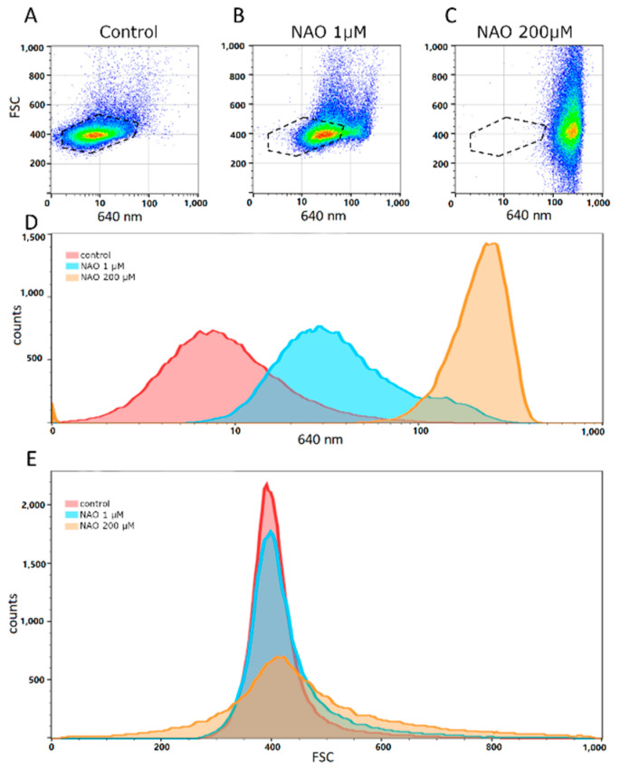
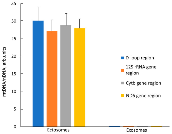
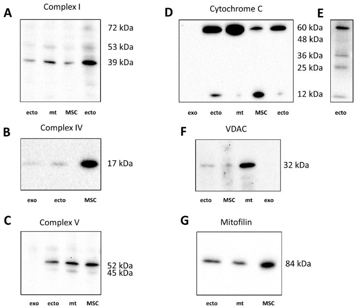
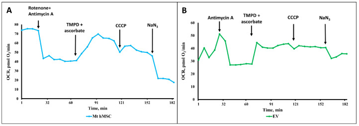
Extracellular vesicles (EV) derived from stem cells have become an effective complement to the use in cell therapy of stem cells themselves, which has led to an explosion of research into the mechanisms of vesicle formation and their action. There is evidence demonstrating the presence of mitochondrial components in EV, but a definitive conclusion about whether EV contains fully functional mitochondria has not yet been made. In this study, two EV fractions derived from mesenchymal stromal stem cells (MSC) and separated by their size were examined. Flow cytometry revealed the presence of mitochondrial lipid components capable of interacting with mitochondrial dyes MitoTracker Green and 10-nonylacridine orange; however, the EV response to the probe for mitochondrial membrane potential was negative. Detailed analysis revealed components from all mitochondria compartments, including house-keeping mitochondria proteins and DNA as well as energy-related proteins such as membrane-localized proteins of complexes I, IV, and V, and soluble proteins from the Krebs cycle. When assessing the functional activity of mitochondria, high variability in oxygen consumption was noted, which was only partially attributed to mitochondrial respiratory activity. Our findings demonstrate that the EV contain all parts of mitochondria; however, their independent functionality inside EV has not been confirmed, which may be due either to the absence of necessary cofactors and/or the EV formation process and, probably the methodology of obtaining EV.

Keywords: ectosomes; exosomes; extracellular vesicles; mesenchymal stromal cells; mitochondria; stem cells.


Conflict of interest statement

The authors declare no conflict of interest.

Figures

Figure 1
Figure 1
(A) Budding of the MSC cell membrane (shown by arrow) preferably in the future forming EV; (B) budding of the membrane with the presence of membranous structures inside (shown by arrow). Note the mitochondria (M) adjacent to the locus; (C) a group of vesicles adjacent to the cell membrane of the MSC; (D) a group of vesicles gathered in the intracellular space of the MSC. Electron microscopy. Scale, 250 nm.
Figure 2
Figure 2
Examples of different configurations of ectosomes obtained by conventional (A) and cryo (B) electron microscopy after centrifugation of the MSC culture medium at 10,000× g. Note the heterogeneity of sizes with average around 250 nm, the frequent presence of shell-like profiles in (A), and the presence of membrane-like structures in the EV lumen in all images in A and in a simple image in (B) shown by a long arrow. Short arrows in (B) demonstrate the presence of vesicles within a multivesicular body. Bar, 250 nm in (A) and 300 nm in (B).
Figure 3
Figure 3
Examples of different configurations of exosomes obtained by conventional (A) and cryo (B) electron microscopy of vesicles supported on grid (grey long profiles) after centrifugation of the MSC culture medium at 108,000× g. Note the heterogeneity of sizes with average around 100 nm. Bar, 150 nm.
Figure 4
Figure 4
The light scattering intensity (A,B) and the numbers of particles (C,D) in two fractions of vesicles derived from MSC estimated by dynamic light scattering measurement using a Zetasizer NanoZS.
Figure 5
Figure 5
EV characterization by NTA analysis obtained in a sample either after 108,000× g centrifugation (exosomes, (A), camera gain = 500; histogram values range: 185–1885, n =12), or ectosomes (B) obtained after centrifugation at 10,000× g of cultivation medium (n =12), camera gain = 225; histogram values range: 65–1560. NTA analysis represents data of mean values with standard error of mean.
Figure 6
Figure 6
Analysis of the mitochondrial probe levels in various types of EV (ectosomes and exosomes) by flow cytometry. ((AC,GI), ectosomes; (DF,J–L), exosomes). Dot-plots of unstained EV (empty vesicles) and EV loaded with MitoTracker Green (MTG, (AF)) and TMRE (GL) are presented. (C,F,I,L) Histograms of the distribution of fluorescent responses for EV loaded with MTG and TMRE on the right side of dot-plots are shown (dark green and dark red, empty EV and light green and light brown are EV loaded with MTG and TMRE correspondingly).
Figure 7
Figure 7
Flow cytometry analysis showing possible cardiolipin presence in ectosomes stained with 10-nonyl-acridine orange (NAO). (AC) Size (y scale, FSC) versus fluorescent intensity (x scale, 640 nm fluorescence signal) dot-plot of control vesicles and those stained with 1 μM and 200 μM NAO. (D) Fluorescence (640 nm) distribution (histogram) in control vesicles and those stained with 1 and 200 μM NAO. Note the presence of a high fluorescent subpopulation in the 1 μM NAO group, which possibly indicates presence of cardiolipin. (E) Size (FSC) distribution in control vesicles, and those stained with 1 μM and 200 μM NAO.
Figure 8
Figure 8
Relative mitochondrial DNA (mtDNA) content in ectosomes and exosomes. The ratio of mtDNA to nuclear DNA (nDNA) in intact MSC has been taken as 1. Four different components of mitochondrial DNA including non-coding region (D-loop) and three different gene regions were examined. Obvious enrichment of ectosomes with mitochondrial DNA (up to 30 fold) and corresponding depletion of mtDNA in exosomes (down to 0.3 fold) is shown.
Figure 9
Figure 9
Western blots of vesicles (ectosomes, ecto and exosomes, exo), MSC, and mitochondria isolated from MSC. MWM with proper molecular masses were calculated based on molecular weight markers. For better representation of different forms of cytochrome C, the separate blot for ectosomes was made with more time exposure of the membrane. In the blot for complex I (with major 39 kDa component) in two separate lanes corresponding to ectosomes the proteins levels were different. For better demonstration of multiple forms, in the rightest lane in (A), the protein content of ectosomes was 4 times higher than in the very left lane (see details in Section 4 and explanations in the text). (AD), probes for Complex I, Complex IV, Complex V and Cytochrome C, correspondingly; (E), example of resolution of bands for Cytochrome C in ectosomes; (F,G) probes for Complex V and Mitofilin.
Figure 10
Figure 10
Oxygen consumption rate (OCR) of mitochondria isolated from MSC ((A), Mt hMSC, blue trace) and ectosomes ((B), EV, green trace). In (A), the medium contained pyruvate (10 mM), malate (2 mM), NADH (1 mM), cytochrome C (5 µM). In (B), the medium contained succinate (10 mM), rotenone (3 µM). Additions in ports: rotenone (3 µM), antimycin A (4 µM), TMPD (150 µM), ascorbate (10 mM), CCCP (1 µM), NaN3 (5 mM).

Similar articles

Cited by

References

    1. Block T.J., Marinkovic M., Tran O.N., Gonzalez A.O., Marshall A., Dean D.D., Chen X.-D. Restoring the Quantity and Quality of Elderly Human Mesenchymal Stem Cells for Autologous Cell-Based Therapies. Stem Cell Res. Ther. 2017;8:239. doi: 10.1186/s13287-017-0688-x. - DOI - PMC - PubMed
    1. Weng Z., Wang Y., Ouchi T., Liu H., Qiao X., Wu C., Zhao Z., Li L., Li B. Mesenchymal Stem/Stromal Cell Senescence: Hallmarks, Mechanisms, and Combating Strategies. Stem Cells Transl. Med. 2022;11:356–371. doi: 10.1093/stcltm/szac004. - DOI - PMC - PubMed
    1. Shimada I.S., Spees J.L. Stem and Progenitor Cells for Neurological Repair: Minor Issues, Major Hurdles, and Exciting Opportunities for Paracrine-Based Therapeutics. J. Cell Biochem. 2011;112:374–380. doi: 10.1002/jcb.22963. - DOI - PMC - PubMed
    1. Yáñez-Mó M., Siljander P.R.-M., Andreu Z., Zavec A.B., Borràs F.E., Buzas E.I., Buzas K., Casal E., Cappello F., Carvalho J., et al. Biological Properties of Extracellular Vesicles and Their Physiological Functions. J. Extracell. Vesicles. 2015;4:27066. doi: 10.3402/jev.v4.27066. - DOI - PMC - PubMed
    1. Budnik V., Ruiz-Cañada C., Wendler F. Extracellular Vesicles Round off Communication in the Nervous System. Nat. Rev. Neurosci. 2016;17:160–172. doi: 10.1038/nrn.2015.29. - DOI - PMC - PubMed

MeSH terms

LinkOut - more resources